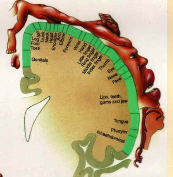
Primary somatosensory cortex
From Psy3241
The Primary Somatosensory Cortex (S1)
Contents |
Purpose:
The areas involved are related to touch. Neurons are activated when the skin is stimulated. The somatosensory cortex acts as the end of the sensory pathway.
Location:
The cerebral cortex in the parietal lobe houses the primary somatosensory cortex (S1). S1 is located behind central sulcus in postcentral gyrus.
History and Basic Anatomy:
Dr. Wilder Penfield was able to construct a map of the human somatosensory cortex in the 1950’s. His strategy was to find a group of patients undergoing brain surgery for epilepsy, stimulate their cortices, and ask the patients what they felt. Through these stimulations, Dr. Penfield was able to observe the parts of the brain that caused the patients to feel sensations on different regions of their bodies. Penfield found that even though one’s arms and trunk make up a lot of the body’s surface area (and, therefore, skin area), not a lot of the primary somatosensory neurons are devoted to these areas. Conversely, the face and hands take up most of the primary somatosensory cortex. This fact makes sense because the amount of cortex devoted to each area is directly proportionate to the density of nerve receptors in those areas. For example, think about how much more sensitive your fingertips are than parts of your legs.
Organization:
The thalamus projects sensory information first to the most rostral part of the S1 (the areas situated more toward the nasal region, known as Brodmann’s areas 3a and 3b). 3a and 3b then relay information and exchange information with Brodmann’s areas 1 and 2.
Brodmann’s area 3b senses primarily texture, size, and shape, Brodmann’s area 1 receives information about texture, and Brodmann’s area 2 receives information about size and shape.
There is a topographical organization (as Penfield found) to the projection of information from the thalamus to the primary somatosensory cortex. A large region close to the base of the postcentral gyrus is devoted to sensations from the tongue, mouth, and lips. Information from the lower limbs and genitals are arranged along the midline. The fingers, lips, and tongue are well represented within the primary somatosensory cortex.
Sources:
http://faculty.washington.edu/chudler/brainsize.html
http://www.neurosci.pharm.utoledo.edu/MBC4420/sensory.htm

