Corpus callosum
From Psy3241
| (11 intermediate revisions not shown) | |||
| Line 3: | Line 3: | ||
The '''corpus callosum''' (Latin for “tough body”) is a broad, thick bundle of nerve fibers in the entire nervous system, running from side to side and consisting of millions and millions of nerve fibers. If we cut a brain in half down the middle, we would also cut through the fibers of the corpus callosum. | The '''corpus callosum''' (Latin for “tough body”) is a broad, thick bundle of nerve fibers in the entire nervous system, running from side to side and consisting of millions and millions of nerve fibers. If we cut a brain in half down the middle, we would also cut through the fibers of the corpus callosum. | ||
| - | + | When looking at the middle side of one half of the brain, in magnetic resonance imaging (MRI), the corpus callosum looks like a section of a mushroom cap located at the center of the brain. [http://www.youtube.com/watch?v=7zOa3LLrHKc Video of Corpus callosum] | |
---- | ---- | ||
| - | |||
[[Image:Col.gif]] | [[Image:Col.gif]] | ||
| - | |||
---- | ---- | ||
| Line 14: | Line 12: | ||
In a healthy infant’s brain, the corpus callosum develops between 12 to 16 weeks after conception. The fibers of the corpus callosum continue to become more and more effective into adolescence. By the time a child is around the age of 12 years, the corpus callosum functions will in adulthood, allowing rapid interaction between the two sides of the brain. From this age on the corpus callosum becomes increasingly functional in their typically developing children. | In a healthy infant’s brain, the corpus callosum develops between 12 to 16 weeks after conception. The fibers of the corpus callosum continue to become more and more effective into adolescence. By the time a child is around the age of 12 years, the corpus callosum functions will in adulthood, allowing rapid interaction between the two sides of the brain. From this age on the corpus callosum becomes increasingly functional in their typically developing children. | ||
---- | ---- | ||
| - | [[Image:Bbb.gif]] | + | == Close-up Sagittal Section of Hemisphere == |
| - | --- | + | [[Image:Bbb.gif]] |
| + | |||
| + | 1.)Rostrum of corpus callosum | ||
| + | |||
| + | 2.)Genu of corpus callosum | ||
| + | |||
| + | 3.)Body of corpus callosum | ||
| + | |||
| + | 4.)Splenium of corpus callosum | ||
| + | |||
| + | 5.)Septum pellucidum | ||
| + | |||
| + | 6.)Anterior commissure | ||
| + | |||
| + | 7.)Fornix | ||
| + | |||
| + | 8.)[[Hippocampus]] | ||
| + | |||
| + | 9.)Cingulate gyrus | ||
| + | |||
| + | 10.)Paraterminal gyrus | ||
| + | |||
| + | == Corpus callosotomy == | ||
| + | |||
| + | Severing the corpus callosum became a method of dealing with and localizing severe epileptic seizures. Sometimes, seizures in the brain will begin in one place and spread throughout the entire brain. The corpus callosum can be severed to prevent seizures from reaching the other hemisphere or, in some cases, to stop the seizures altogether. This procedure is known as a ''corpus callosotomy'' and results in the split-brain condition. | ||
| + | |||
| + | |||
| + | The split-brain condition is where hemispheres of the brain can no longer interact with each other due to the severing of the corpus callosum. For example, when a split-brain patient is shown an object, such as a cup, in their left visual field, they will be unable to name it. This is because the visual stimulus enters the right hemisphere through the left eye, but the information can not travel to the left hemisphere to be recognized by the language center of the brain. Thus it is impossible for the subject to name the object unless they are able to turn their head and move the object into their right visual field. However, if they are asked to identify the object using a pencil in their left hand to draw or write with, they can identify the object. | ||
== References == | == References == | ||
| + | The Anatomy Project, (2000). Close-up Sagittal Section of Hemisphere. Retrieved April 19, 2008, from Medical Gross Anatomy Web site: http://anatomy.med.umich.edu/atlas/n1a5p8.html | ||
| + | |||
Hubel, D. H (2006). THE CORPUS CALLOSUM. Retrieved April 21, 2008, from Eye, Brain, and Vision Web site: http://hubel.med.harvard.edu/b34.htm | Hubel, D. H (2006). THE CORPUS CALLOSUM. Retrieved April 21, 2008, from Eye, Brain, and Vision Web site: http://hubel.med.harvard.edu/b34.htm | ||
Current revision as of 07:28, 25 April 2008
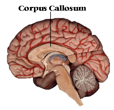
The corpus callosum (Latin for “tough body”) is a broad, thick bundle of nerve fibers in the entire nervous system, running from side to side and consisting of millions and millions of nerve fibers. If we cut a brain in half down the middle, we would also cut through the fibers of the corpus callosum.
When looking at the middle side of one half of the brain, in magnetic resonance imaging (MRI), the corpus callosum looks like a section of a mushroom cap located at the center of the brain. Video of Corpus callosum
Contents |
Development
In a healthy infant’s brain, the corpus callosum develops between 12 to 16 weeks after conception. The fibers of the corpus callosum continue to become more and more effective into adolescence. By the time a child is around the age of 12 years, the corpus callosum functions will in adulthood, allowing rapid interaction between the two sides of the brain. From this age on the corpus callosum becomes increasingly functional in their typically developing children.
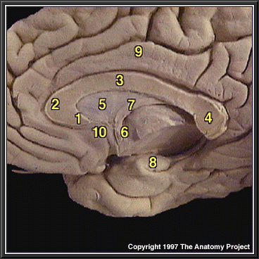
Close-up Sagittal Section of Hemisphere
1.)Rostrum of corpus callosum
2.)Genu of corpus callosum
3.)Body of corpus callosum
4.)Splenium of corpus callosum
5.)Septum pellucidum
6.)Anterior commissure
7.)Fornix
8.)Hippocampus
9.)Cingulate gyrus
10.)Paraterminal gyrus
Corpus callosotomy
Severing the corpus callosum became a method of dealing with and localizing severe epileptic seizures. Sometimes, seizures in the brain will begin in one place and spread throughout the entire brain. The corpus callosum can be severed to prevent seizures from reaching the other hemisphere or, in some cases, to stop the seizures altogether. This procedure is known as a corpus callosotomy and results in the split-brain condition.
The split-brain condition is where hemispheres of the brain can no longer interact with each other due to the severing of the corpus callosum. For example, when a split-brain patient is shown an object, such as a cup, in their left visual field, they will be unable to name it. This is because the visual stimulus enters the right hemisphere through the left eye, but the information can not travel to the left hemisphere to be recognized by the language center of the brain. Thus it is impossible for the subject to name the object unless they are able to turn their head and move the object into their right visual field. However, if they are asked to identify the object using a pencil in their left hand to draw or write with, they can identify the object.
References
The Anatomy Project, (2000). Close-up Sagittal Section of Hemisphere. Retrieved April 19, 2008, from Medical Gross Anatomy Web site: http://anatomy.med.umich.edu/atlas/n1a5p8.html
Hubel, D. H (2006). THE CORPUS CALLOSUM. Retrieved April 21, 2008, from Eye, Brain, and Vision Web site: http://hubel.med.harvard.edu/b34.htm
NODCC, (2006). What is the Corpus Callosum? . Retrieved April 21, 2008, from National Organization for Disorders in the CorpusCallosum Web site: http://www.nodcc.org/what_is_the_corpus_callosum.php


