Dermatology - Scaling Disorders
From Iusmicm
Contents |
[edit] Scaling Disorders
- Scaling disorders are called papulosquamous: something you can feel + scaling
- Scale comes from the French word escale and the English skale which mean a substance that separates like a husk.
- Rash comes from the French word rache meaning "eruption" and the Latin radere meaning "sracth" indicating the sudden appearance and severity of a rash.
- Scaling rashes usually generate excess keratinocytes.
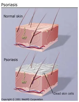
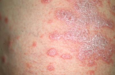
[edit] Psoriasis
- The prototypical scaling rash
- Biblical leprosy may actually be referencing psoriasis
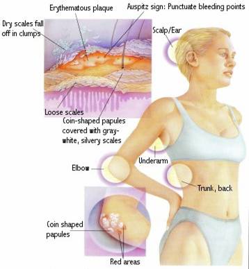
- Cutaneous characteristics:
- Well-defined erythematous papules and plaques with silvery scaling
- Distribution: symmetrical, at extensor surfaces (elbows and knees), buttocks
- Erythroderma (abnormal redness); "red man syndrome"
- Can be so bad they have a hard time maintaining temperature and water balance
- Redness almost always followed by peeling
- 7-fold faster epidermal production (every 3-4 days), thus a thickening
- Common sites: extensor surfaces (elbows, knees), sacrum, trauma sites (Koebner phenomenon), scalp, heel


- Histological characteristics:
- Inflammation
- Parakeratosis (retention of nuclei in keratinocytes)
- Acanthosis (thickening of the epidermis)=


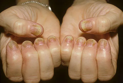
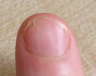
- Non-cutaneous characteristics:
- Nail involvement: pits, onycholysis (nail separation)
- Lots of nail pits indicates likely joint issues, too.
- Arthritis in 5% of pts
- Increased risk for metabolic disease and cardiac-related infections
- Nail involvement: pits, onycholysis (nail separation)


- Clinical characteristics:
- Scales may be minimal or masked by hydration
- Onset can be at any age.
- Has a genetic predisposition: family history positive in 1/3 of cases
- Can be precipitated by: infection, trauma, or drugs.
- Can actually IMPROVE with sun exposure.
- Koebner phenomenon describes how lesions tend to occur at sites of trauma.
- Often arises after streptococcal pharyngitis; think superantigens
- Note that potent topical steroid treatment leads to permanent stria!
- Be careful where you apply topical steroids: don't use it on genitals or faces
- PUBA sensitizes cells to ultraviolet and kills the excessively dividing cells
- Psoriasis subtypes:
- Acute onset Guttate psoriasis
- Guttate = teardrop in shape
- "Guttate psoriasis is characterized by teardrop-size pink papules that often develop in response to a streptococcal or other upper respiratory tract infection. The lesions are much smaller than those of psoriasis vulgaris; however, this usually shortlived condition can evolve into chronic psoriatic disease." per ConsultLive
- Associated with strep infection


- Perineal psoriasis
- Acute onset Guttate psoriasis

- Other images of psoriasis:
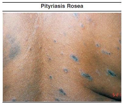
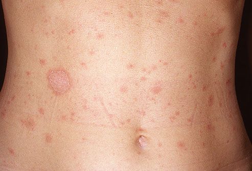
[edit] Pityriasis rosea
- Cutaneous characteristics of pityriasis rosea:
- Comes in several forms of color, shape, and lesion type:
- Shape: round OR oval
- Color: pink OR brown
- Lesion type: macules / patches (non-palpable, discolored) OR papules / plaques (raised)
- Herald patch: heralds the coming of the infection
- Much thinner than psoriasis
- No nail involvement with PR
- Papular version (Herald patches): more inflammatory version; red, irregular, papules with a scale within the erythmatous border
- Classic presentation: red-yellow, oval / round, (thin) plaques with trailing scale; oriented along cleavage lines
- Symmetry: distributed over both sides of the trunk
- Distribution: primarily truncal; follows skin lines; "oriented along cleavage lines"
- Mild puritis


- Comes in several forms of color, shape, and lesion type:
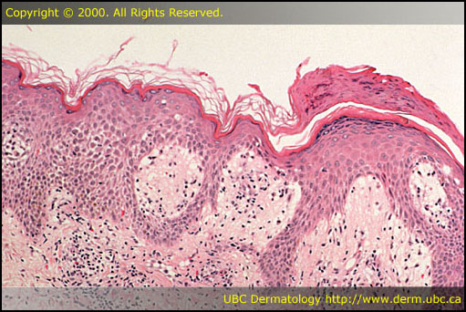
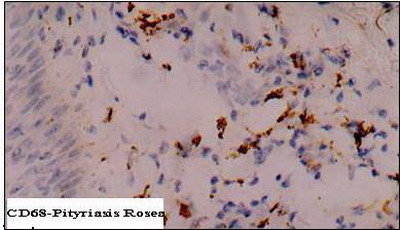
- Histological characteristics of pityriasis rosea:
- Clinical characteristics of pityriasis rosea:
- Acute, self-limiting
- Unknown cause, though probably herpes
- Weird pattern of spread: entire classroom yet not within a family
- Consider secondary syphillis in the differential of pityriasis rosea
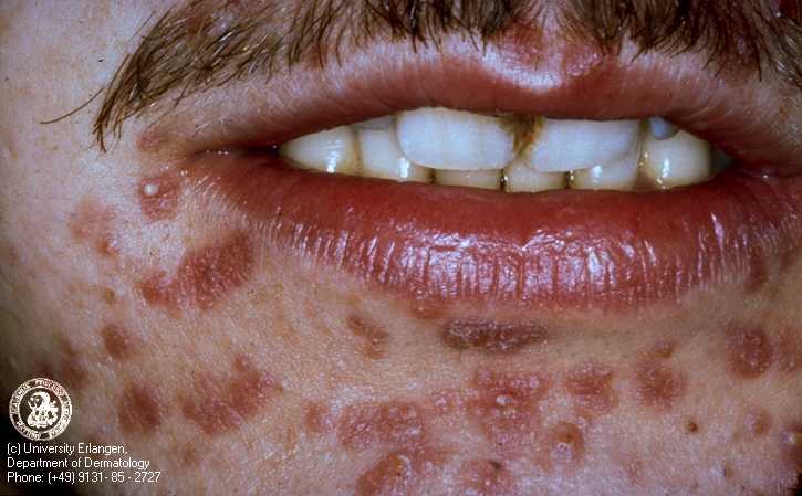
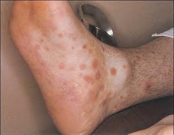
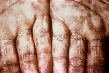
[edit] Secondary syphillis
- Cutaneous characteristics:
- Chancres!
- Will go away on its own, often
- Copper pennies with scaling
- More brown-ish than red
- Teeming with spirochetes
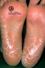
- On palms or soles
- Distribution: mouth, genitalia / anus (condyloma lata), palms of hands, planar surface of feet, back,
- Condyloma lata look like warts but won't respond to wart tx.


- http://infections.consultantlive.com/image/image_gallery?img_id=1400637&t=1239393788816
- http://infections.consultantlive.com/image/image_gallery?img_id=1400629&t=1239628194012
- http://infections.consultantlive.com/image/image_gallery?img_id=1400641&t=1239393788843
- http://infections.consultantlive.com/image/image_gallery?img_id=1400645&t=1239393788865
- http://infections.consultantlive.com/image/image_gallery?img_id=1400653&t=1239393788926
- http://infections.consultantlive.com/image/image_gallery?img_id=1400657&t=1239628194124


- Chancres!
- Non-cutaneous characteristics of secondary syphillis (a systemic disease):
- Fever
- Mucous membranes
- Headache
- Enlarged lymph nodes
- Clinical characteristics of secondary syphillis:
- A systemic disease!
- If you suspect syphillis put on gloves!
- Blood tests are nearly 100% sensitive / specific so if you get a positive, they have it.
- 1/3 of untreated cases will resolve on their own without incident
- Other images of secondary syphillis:

[edit] Lichen planus
- Cutaneous characteristics of lichen planus:
- Pruritus: really, really, really itchy!
- Four P's: purple, polygonal, pruritic, papules / plaques.
- Look like lichen ("A simple slow-growing plant that typically forms a low crustlike, leaflike, or branching growth on rocks, walls, and trees" per Google)
- An inflammatory cutaneous and mucosal membrane disease
- 'Flat topped papules / plaques that are violaceous (violet in color, "more purple than expected") and scaling
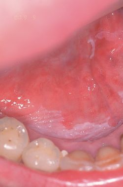
- Key word is Whickham striae which are the white lacy patterns.
- These can look like psoriasis but they are purple not red.
- Distribution: trunk and extremities, especially the wrists and distal leg, oral mucosa
- Note that if the oral lacy formations don't scrape off, it isn't candida and it is likely lichen planus
- Can present as similar to graft-versus-host disease'
- Demonstrates "Koebner phenomenon" (occurring near sites of trauma / scratching)
- Results in linear distribution when due to scratching
- Recall that psoriasis, too, demonstrates Koebner phenomenon


-


- Clinical characteristics of lichen planus:
- Unknown cause (like pityriasis rosea)
- Not contagious
- Abrupt onset
- Skin and mucosal membrane involvement
- Hard to treat
- Can include the nail
[edit] Chronic dermatitis: Atopic dermatitis
- Cutaneous characteristics of atopic dermatitis:
- Erythema with lichenification, excoriations, crusting, and pruritis
- Lichenification: looks like a washboard and requires 200k sratches
- Pruritis
- Distribution: flexor surfaces (popliteal fossa, cubital fossa, posterior cervical region), cheeks


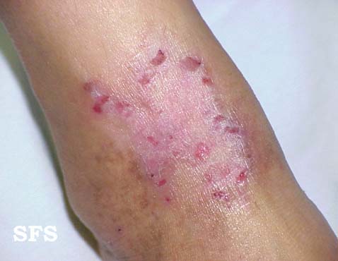


- Erythema with lichenification, excoriations, crusting, and pruritis
- Non-cutaneous characteristics of atopic dermatitis:

- Clinical characteristics of atopic dermatitis:
- Associated with asthma / hayfever
- Family history is important
- Elevated IgE levels
- "The itch that rashs"
- Chronic results in thickening
- Sub-acute results in crusting, oozing, dryness
[edit] Chronic dermatitis: Seborrheic dermatitis
- Cutaneous characteristics of seborrheic dermatitis:
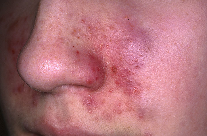
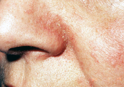
- Erythematous papules characterized by greasiness and scaling
- Distribution: symmetrical; sternum, axilla, scalp, naso-oral area, medial to scapulae, behind the ear
- Characterized by increases sebaceous gland production of oil ("greasiness")
- Infant form is called "cradle cap"


- Non-cutaneous characteristics of seberrheic dermatitis:
- HIV is a risk factor
- Parkinsons results more frequently
- Clinical characteristics of seborrheic dermatitis:
- Unknown cause
- Pityrosporium species may be involved
- Elevated frequency in AIDS and Parkinson disease
- Other images of seborrheic dermatitis:


[edit] Chronic dermatitis: Stasis dermatitis
- Cutaneous characteristics of stasis dermatitis:
- Erythematous, brown lesions with a sharp border, scaling, ulcerations, and crusting
- Think ulcers; very difficult to treat
- Edema
- Fibrosis

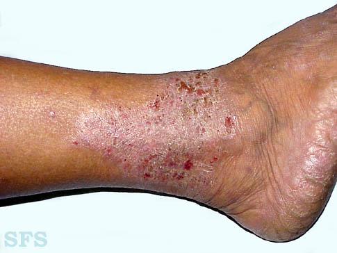
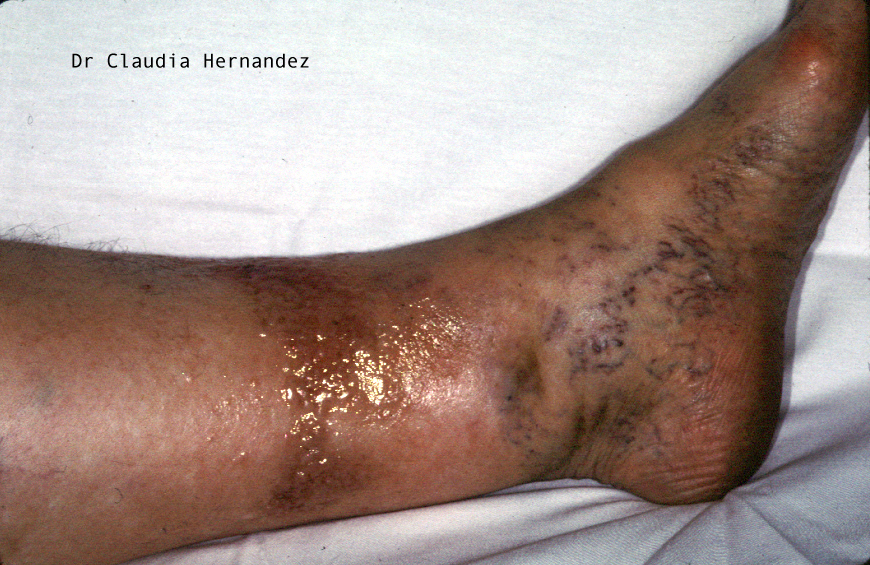

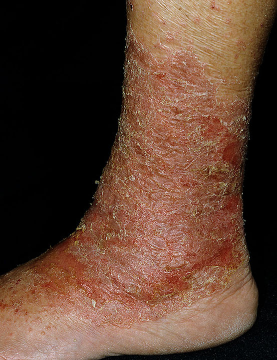

- Non-cutaneous characteristics of stasis dermatitis:
- Peripheral venous disease
- Hemosiderin abnormalities
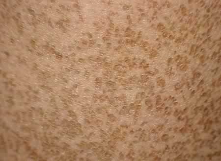
[edit] Ichthyosis
- Cutaneous characteristics:
- Fish skin with scaling, no erythema (lack of inflammation), and a white or brown coloration
- Distribution: pectoral region, palms (hyperlinear), keratosis pilaris (rough bumps on the skin from an auto-dominant follicular disorder)


- Non-cutaneous characteristics:
- Potential for cancers
- Clinical characteristics of ichthyosis:
- Genetics play an important role
- Recall that keratosis pilaris is seen which is an auto-dominant follicular disorder
- Genetics play an important role
**Associated with internal disease
[edit] Superficial fungal infections
- Cutaneous characteristics of fungal infections:
- Erythematous macules / patches with scaling
- Tinea capitis / corporis
