Dermatology - Cutaneous Signs of Systemic Disease
From Iusmicm
Contents |
[edit] Cutaneous Signs of Systemic Disease
[edit] Rheumatology
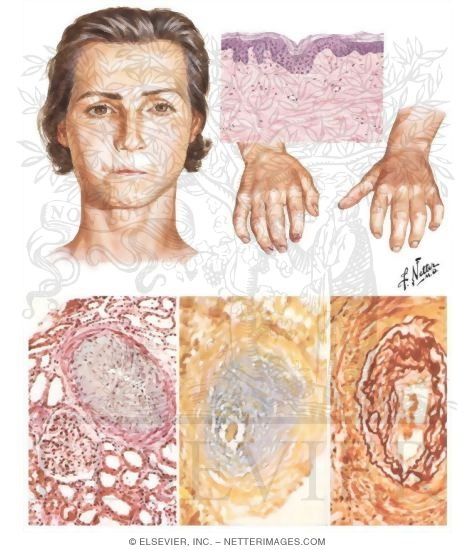
[edit] Lupus erythematosus
[edit] Acute lupus erythematosus
- Cutaneous symptoms:
- NB: lupus erythematous lesions are blanchable
- Distribution is along photon exposure
- Look for a malar rash
- Violaceous = purple lesion; often means there are lymphocytes in the epidermis.
- Systemic symptoms:
- circulating antibodies
- immunoglobulin deposition in skin and other organs

[edit] Chronic (discoid) lupus erythematosus
- Cutaneous symptoms:
- Erythema with telangiectasis
- Scaling, follicular plugging
- Scarring that leads to alopecia
- Hypopigmentation on the inside and hyperpigmentation on the outside.


- Systemic symptoms:
- Discoid LE usually remains localized to the skin
- 5% will develop systemic symptoms like immunoglobulin
[edit] Dermatomyositis
- Cutaneous symptoms:
- Gottron's papules over joints
- Periungual erythema and "shaggy" cuticles
- Periorbital heliotrope eruption: erythema / violaceous around the eyes
- Spares the upper lip

[edit] Scleroderma
- Cutaneous symptoms:
- Called "localized scleroderma"
- Light macules / papules
- Hard to see; feels like hard scar tissue
- Shows atrophy of the epidermis
- Scarring alopecia
**Follows a dermatome?


- Systemic symptoms:
- Usually started in hands
- Rounded off digits
- Increased deposition of collage in the heart / lungs / kidneys / GI tract / joints / skin

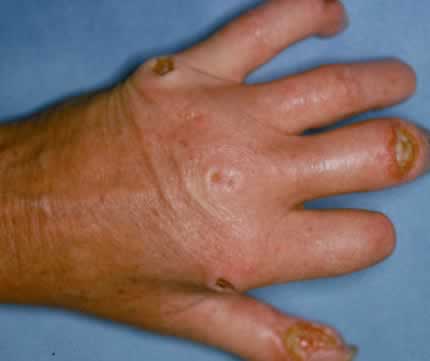
[edit] Vasculitis
- Cutaneous symptoms:
- Palpable purpura
**Biopsy to diagnose vasculitis

- Systemic symptoms:
- Henoch-Schonlein purpura associated with renal or bowel involvement
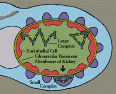
- Type 3 Ab reaction (immune complex) against the endothelium of the vasculature
- Antibodies bind to an antigen and form a complex
- The complex activates platelets (clotting) and the complement pathway (inflammation)
- C3a and C5a are important complement factors that lead to inflammation
- Vasoactive amines increase permeability and lead to erythema
- Microthrombi are formed because of the activation of clotting on the deposits of immune complexes.
- Endothelial damage occurs because of the inflammation that leads to enzyme release (neuts, basophils, etc.)


- Hypersensitivity vasculitis:
- Has 5 criteria; 3 or more required for dx:
- Age >16
- Use of a possible offending drug in temporal relation to the symptoms
- Palpable purpura
- Maculopapular rash
- Biopsy of a skin lesion showing neutrophils around an arteriole or venule

[edit] Aphthous ulcer
- Come in minor and major forms
- Little eronsions on the oral mucosa
- Minor are not usually a sign of systemic disease
- Think LE and HIV


[edit] Behcet's
- Results in erythema nodosum
- Think HIV
- Erythema nodosum: type 3 hypersensitivity in the fat tissue

[edit] Endocrinology
[edit] Hypothyroidism (Myxedema)
- Myxedema: "Swelling of the skin and underlying tissues giving a waxy consistency, typical of patients with underactive thyroid glands"
- Lateral eyebrow loss in myxedema is classic board question.
- Mucopolysaccharides in the skin.
- Cutaneous symptoms:
- Puffy skin
- Yellowish tint
- Loss of lateral eyebrows
- Dry, course, brittle hair

- Systemic symptoms:
- Typical hypothyroid stuff
[edit] Hirsutism
- Excess androgen production
- Can be from ovary, adrenals, or drug-induced
- Mind the racial differences and normal variation
- Cutaneous symptoms:
- Lots of hair

- Systemic symptoms:
- may be an endocrine tumor
[edit] Diabetes Mellitus
- Cutaneous symptoms:
- Necrobiosis lipoidica: well-demarcated areas of epidermal atrophy
- Yellowing ulcerations
- Telangiectasias
- Anterior lower legs


- Necrobiosis lipoidica: well-demarcated areas of epidermal atrophy
[edit] Pulmonary
[edit] Sarcoidosis
- Cutaneous symptoms:
- Nasal / facial papules / plaques
- Fingernail infections
- Intra-scapular papules


- Systemic symptoms:
- Think lung issues
- Distal digit bone deterioration

[edit] Gastroenterology
[edit] Metabolic Disorders
[edit] Hyperlipidemia
- Cutaneous symptoms:
- Xanthomas: firm, flesh-colored to yellowish, papules / plaques
- At the eye, a xanthoma is called a xanthelasma
- Xanthelasmas usually DO NOT indicate an underlying lipid issue


- Xanthomas: firm, flesh-colored to yellowish, papules / plaques
[edit] Porphyria cutanea tarda
- Cutaneous symptoms:
- Blisters / bullae and scarring on dorsum of hand
- Hypertrichosis
- Can be dx by demonstrating fluorescent serum


- Systemic issues:
- Liver disease
- Acquired porphyria
[edit] Chronic liver diasease
- Cutaneous symptoms:
- Spider telangiectasias
- Palmar erythema
- Jaundice

[edit] Inflammatory bowel disease
- Cutaneous symptoms:
- Ulcerations characterized by their rolled violaceous border
- Called pyoderma gangrenosum: 50% have an underlying disease (think IBD, LE)
- DO NOT DEBRIDE!


[edit] Hematology / Oncology
[edit] Leukemia
- Cutaneous symptoms:
- Leukemia cutis: purpuric or flesh-colored papules and plaques
- Characteristically round
- 10-50% of monocytic leukemia pts manifest leukemia cutis
- 6-20% of lymphocytic and granulocytic leukemias manifest leukemia cutis


[edit] Lymphoma
- Cutaneous symptoms:
- Lymphoma cutis: cutaneous collections of T cells
- appears as macules / pathces, plaques, OR nodules
- Can be present for years, then start evolving into plaques and nodules.
- Mycosis fungoides type...?

- Lymphoma cutis: cutaneous collections of T cells
[edit] Bone marrow transplantation
- Cutaneous symptoms:
- GVHD:
- Acute: Light erythema macules that spread down the head along the cheeks
- Chronic: pink macules at the joints / tips of hands, depigmentation plaques

- GVHD:
[edit] Psychiatry
[edit] Factitial
- Cutaneous symptoms:
- Look for odd borders, weird patterns, strange distributions, et cetera.


-
[edit] Neurotic excoriations
- Cutaneous symptoms:
- Excoriations made by pt's own manipulation: an uncontrolled cycle of re-damaging the skin.

[edit] Cases
[edit] Case 1


[edit] Case 2


- Discoid lupus erythematosus
[edit] Casse 3

- Spider telangiectasias
- Jaundice
- Palmar erythema
- Liver failure
