Cardiology - Aortic Valve Disease
From Iusmicm
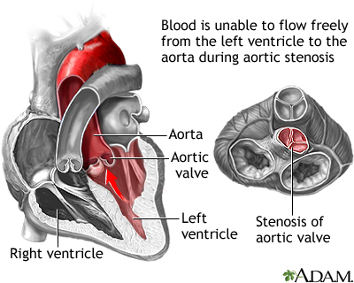
[edit] Aortic Stenosis
- Recall that "stenosis" is a narrowing.
- Sclerosis is a hardening (decrease in compliance).

[edit] A Case
- 67 yo woman referred for murmur which she has known about for a ‘few years.’
- Over recent months she has noticed dyspnea climbing stairs– before she was completely asymptomatic.
- On exam, BP 140/86. JVP appears normal. Carotid upstroke is mildly diminished and delayed. Chest clear to auscultation. Cardiac exam with III/VI systolic ejection murmur at RUSB radiating to carotids and apex
which is late peaking. No diastolic murmur is heard. The PMI is sustained and a soft S4 is heard at the apex.
- Why is the carotid upstroke delayed and diminished?
- What is the underlying valvular pathology and how severe is it?
- What should be done about it?
[edit] Aortic Stenosis: Underlying Causes
- Aortic stenosis can result from four underlying causes: calcification, rheumatic disease, congenital malformations (bicuspid, unicuspid), and radiation treatment.
- For pts < 70 yo, the major causes of aortic stenosis are bicuspid, rheumatic, and calcification.
- For pts > 70 yo, the major causes of aortic stenosis are calcification, bicuspid, and rheumatic.

[edit] Aortic Valve Sclerosis
- Recall that in general, sclerosis is a decrease in compliance due to thickening or scarring.
- Specifically we say "sclerosis is a focal thickening of valve cusps with normal excursion and no obstruction to flow".
- The pathophysiology of a sclerotic valve is similar to atherosclerosis (think plaques, ulcerations, clotting, etc.)
- Aortic valve sclerosis is a normal development with age as 25% of pts over 65 yo have a sclerotic aortic valve.
- NB: aortic valve sclerosis may progress to aortic stenosis.
- In addition, sclerotic aortic valves double the risk of coronary events.
- Sclerotic aortic valves generate an early peaking systolic ejection murmur (SEM) at the right upper sternal border (RUSB).
- This murmur is a function of normal flow over abnormal anatomy.
- The longer the systolic murmer (decrescendoing), the worse the stenosis


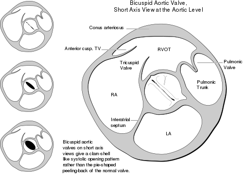
[edit] The Bicuspid Aortic Valve
- Bicuspid aortic valves are found in 1-2% of the population.
- Bicuspid aortic valves are 4 times more common in males.
- Like aortic valve sclerosis, a bicuspid valve can lead to stenosis.
- In addition, an aortic bicuspid valve can produce regurgitation.
- Indeed, many pts with aortic bicuspid valve will require surgery.
- Aortic bicuspid valves moderately increase the risk of endocarditis.

- The bicuspid aortic valve will produce a systolic ejection click as the two leaflets catch on each other upon opening.
- Bicuspid aortic valve (an therefore a systolic ejection click) should be a hint to check for aortic disorders:
- Ascending aortic aneurysm in 50% of pts with a bicuspid aortic valve
- Coarctation in 10% of pts with a bicuspid valve
- 30-50% with coarctation have a bicuspid aortic valve
- Aortic dissection in pts with a bicuspid aortic valve


�===Aortic Stenosis: Pathophysiology===
- The primary pathophysiology behind aortic stenosis is an elevated pressure in the left ventricle as it tries to force the same amount of blood through a smaller (stenotic) hole (aortic valve).
- An elevated pressure in the left ventricle is compensated by left ventricular hypertrophy which makes the left ventricle stronger so it can push the same volume of blood through a stenotic aortic valve.
- This type of compensation is concentric compensation because the left ventricle hypertrophies concentrically by laying down more contracile units concentrically (that is, parallel to) the exisiting units.
- Concentric hypertrophy works initially but with increasing stenosis (and therefore increasing hypertrophy) the cardiac output becomes fixed meaning the pt can no longer respond to increased effort or increased venous return by expanding the ventricle (and therefore increasing output by Starling's principle).
- Concentric hypertrophy fails as stenosis keeps rising but cardiac output declines passed an ability to sustain the pt.
[edit] The Law of LaPlace
- The law of LaPlace is about wall stress because the heart wants to keep wall stress low.

- LaPlace determined that wall stress is a function of pressure, radius, and thickness.
- More pressure, more stress.
- Larger radius, larger stress.
- More thicknes, less stress.
- Wall stress = (Pressure * Radius) / (2 * Thickness)
- Note that the Law of LaPlace applies to alveoli and vasculature, too.


[edit] Aortic Stenosis: Presenting Symptoms
- The signs and symptoms of aortic stenosis are angina, syncope, and heart failure.
- Angina makes sense in the setting of aortic valve stenosis because:
- Muscle mass is increasing (LV hypertrophy b/c must get same volume through stenotic aortic valve) and the heart muscle tissue elicits pain (angina) upon lack of oxygen.
- Coronary flow is reduced (because LV is having a hard time getting blood through aortic valve and also because there is increased resistance in vessels of LV as the muscle has hypertrophied) and the heart muscle tissue elicits pain (angina) upon lack of oxygen.
- There is usually (50% of the time) concurrent coronary artery disease (blockages in the coronary arteries) and the heart muscle tissue elicits pain (angina) upon lack of oxygen.
- Syncope makes sense in the setting of aortic valve stenosis because:
- The heart (left ventricle) can no longer increase output (because of it's concentric hypertrophy and therefore fixed output) to meet increased physical demands so the brain says "I don't have enough oxygen to work with" and shuts down.
- The brain responds to high left ventricular pressure by inducing syncope.
why?
- Heart Failure makes sense in the setting of aortic valve stenosis because:
- The left ventricle has hypertropied (concentrically) to the point of diastolic dysfunction because it is so thick (and therefore stiff).
- The heart (left ventricle) can no longer increase it's output (because concentric hypertrophy has encroached on lumenal space) such that the cardiac tissue itself begins to function with too little oxygen.
- Cardiac tissue develops systolic dysfunction because of a decreasing cardiac output (yet increasing or stable stenosis).
[edit] Aortic Stenosis: Physical Exam
- Recall that aortic stenosis is a narrowing of the LV-aortic junction.
- Recall that aortic stenosis murmurs are due to normal flow through abnormal anatomy (in this case narrowed anatomy).
- Recall that aortic stenosis can result from aortic bicuspid valve, calcification, sclerosis, and rheumatic fever.
- The physical exam findings of aortic stenosis include:
- a systolic ejection click (if stenosis is secondary to a bicuspid valve)
- an early peaking systolic ejection murmur at the RUSB that radiates to the carotids
- This is a crescendo-decrescendo shaped murmur.
- The later in systole the peak intensity, the more severe the stenosis.
- The systolic ejection murmur of aortic stenosis will be "harsh and grade II+".
- a weak and late (parvus (little) et tardus (late)) carotid pulse
- It makes sense that the carotid pulse will be weak and late because with stenosis the blood arrives at the carotid late and with decreased pressure (imagine the small whole zapping some of the normal pressure that would produce the carotid pulse).
- a sustained left ventricle apical impulse
What is apical impulse?
- a fourth heart sound (S4, due to ventricular stiffness because of concentric hypertrophy)
- (S3 may or may not be heard late in the course of aortic stenosis)
- (A2 of S2 may be decreased in severe cases).
- A decreased A2 makes sense as the aortic valve opening becomes very small the valve doesn't have far to close so the closing sound will decrease.
[edit] Aortic Stenosis: Diagnosis
- Diagnosis of aortic stenosis is ultimately by echocardiography.
- Remember, however, that the pt should have the typical physical exam findings: crescendo-decrescendo systolic murmur, parvus et tardus carotid pulse, sustained apical impulse, and S4 (and S3 and decreased A2 in late course).
- The pt should have classic symptoms: angina, syncope, and heart failure.
- Diagnosis may also be aided by observing changes in EKG tracings and cardiac catheterization with pressure differential measured between LV and aorta.
- Recall that the primary pathophysiology of aortic stenosis is increasing pressure in the left ventricle due to decreased output tract area (stenosis).
[edit] Aortic Stenosis: Grading Severity
- Aortic stenosis is measured by the area of the junction between the left ventricle and the aorta and the pressure gradient between the LV and the aorta.
- The smaller the area, the worse (mild -> moderate -> severe).
- The larger the pressure difference, the worse (mild -> moderate -> severe).
[edit] Aortic Stenosis: Natural History
- Recall that aortic stenosis is a narrowing of the LV output tract that causes an increase in LV pressure.
- Recall that the LV undergoes concentric hypertrophy to compensate for the need for increased pressure (to get the volume through the smaller area).
- Recall that concentric hypertrophy eventually decreases the cardiac output causing symptoms.
- Recall that symptoms of aortic stenosis include syncope, angina, and heart failure.
- This process doesn't really stop and the natural history leads to a rapid progression as cardiac output decreases to a point that damages the heart tissue (which cyclically and therefore rapidly leads to worse output and more damage).
- 27-79% of pts with (mild-moderate) aortic stenosis progress ot death or surgery within 5 years.


[edit] Aortic Stenosis: Therapy
- The definitive treatment for aortic stenosis is aortic valve replacement.
- However, percutaneous aortic valvuloplasty is of limited utility in adults
- TAVI = transcatheter aortic valve implantation.
- TAVI is the up and coming treatment.
- TAVI has been shown to be as good as but not better than percutaneous valve replacement.
[edit] Therapy: Indications for AVR (aortic valve replacement)
- Pts are recommended for valve replacement based on their symptoms, LV function, and opportunity.
- Severe symptoms justify replacement.
- LV dysfunction (as defined as ejection fraction (EF) < 50%) justifies replacement.
- Another cardiac surgery provides an opportunity and justification to replace the valve.
- Think CABG, aorta, or mitral valve surgery.
[edit] Aortic Regurgitation
[edit] Aortic Regurgitation: Etiology
- There are two general causes of aortic regurgitation: abnormal valve cusps or abnormal tissue around the valve.
- Tissue surrounding the aortic valve can be damaged by dilitation of the aorta or of the aortic annulus.
- Recall that type A aortic aneurysms can damage the annulus.
- Recall that ascending aortic aneurysms can damage the annulus and cusps
- Ascending aortic aneurysms are associated with connective tissue diseases (Marfan's, Ehlers Danlos, osteogenesis imperfecta), malformations (bicuspid aortic valve, annuloaortic ectasia), and ankylosing spondylitis.
- Recall that a series of artery inflammatory processes can also damage the aorta and annulus: Takayasu's, giant cell, and syphillis.
- Abnormal aortic cusps can also cause aortic regurgitation
- Structural abnormality of the cusp can be genetic (bicuspid), infectious (rheumatic, endocarditis), metabolic (calcific, anorexigens), or iatrogenic (radiation).
- A supracristal ventral septal defect can also cause prolapse of the valve and thus regurgitation.
[edit] Chronic Aortic Regurgitation: Pathophysiology
- So how does the heart respond to aortic regurgitation?
- Blood leaks back into the ventricle during diastole so the end diastolic volume is elevated.
- Because the EDV of the left ventricle is elevated, the heart undergoes eccentric hypertrophy (that is, more myofibrils end-on-end) to make the chamber larger.
- In order to maintain forward flow (because some is leaking back into the ventricle), stroke volume is increased (via concentric hypertrophy).
- Increased stroke volume leads to systolic hypertension and therefore pressure overload of the left ventricle.
- Systolic hypertension leads to more concentric hypertrophy.
- Ultimately, regurgitation gets worse causing worsening volume overload, hypertension gets worse causing worsening pressure overload, eccentric and concentric hypertrophy get more severe, and cardiac output fails.
- The ultimate fate of aortic regurgitation is decreased cardiac output and death.
[edit] Aortic Regurgitation: Law of LaPlace
- As a rule, the heart is trying to maintain the lowest wall stress it can, while still perfusing the body.
- Wall Stress = (Pressure within the ventricle X the Radius) / (2 X Thickness of the ventricle wall)
- Stress = (P X R) / (2 X Th)
- The eccentric hypertrophy of aortic regurgitation is an effort of the heart to reduce the pressure (by dilating the chamber and thus increasing R).
- The concentric hypertrophy of aortic regurgitation is an effort of the heart to maintain cardiac output (by increasing ejection fraction) but this increases thickness.
- This doesn't actually make much sense....
[edit] Aortic Regurgitation: Presenting Symptoms
- Give the story of eccentric and concentric hypertrophy of aortic regurgitation, the symptoms make sense:
- A forceful heart beat because the dilated, thick walled heart is working very hard to force blood into the systemic circulation.
- Angina because the heart is working very hard but much of the blood that should be going into the coronary arteries is draining back into the ventricle.
- Angina is worse when there is concurrent coronary artery disease, when there is low aortic diastolic pressure (worse regurgitation), and when there is more cardiac need for oxygen (worse hypertrophy, more contractility).
- Congestive heart failure because the heart is working hard but there is poor perfusion via the coronary arteries.
- CHF is marked by left ventricle systolic dysfunction, hypertrophy, and an inability to augment cardiac output.
[edit] Chronic Aortic Regurgitation: Physical Exam
- The primary physical exam signs are:
- Wide pulse pressure
- Soft S1
- Systolic ejection click (if regurg due to bicuspid valve)
- Lateral and dynamic PMI displacement
- S3 upon systolic dysfunction
- Immediate diastolic murmur.
[edit] Chronic Aortic Regurgitaiton: Wide Pulse Pressure
- Aortic regurgitation results in wide pulse pressure (that is a >10 mmHg difference in systolic and diastolic pressures) because the heart is working hard (high systolic) and there is much back flow (low diastolic).
- Wide pulse pressures manifest as:
- A brisk / bounding carotid pulse (Corrigan’s pulse)
- Quincke’s pulse (pulsations in nailbeds)
- deMusset’s sign (head bobbing)
- Duroziez’s sign (systolic-diastolic bruit when femoral artery compressed lightly)
- Traube’s sign (‘pistol shot’ sounds over femoral artery)
[edit] Chronic Aortic Regurgitation: Heart Sounds
- An immediate diasolic murmur is present upon aortic regurgitation (which makes sense as the elastic aorta squirts blood back into the ventricle).
- There is a soft S1 (which makes sense as the aorta won't really close nicely).
- There may be an S3 present if there is LV systolic dysfunction.
- An ejection murmur may also be present.
- The ejection murmur will be characterized as immediate, high-pitched, and decrescendo.
- The ejection murmur will be heard at the 3rd left intercostal space, leaning forward, with held expiration.
- The murmur may be heard at the 2nd right intercostal if there is aortic root dissection causing the aortic regurgitation.
- The LONGER the murmur the more severe the regurgitation.
- The louder the murmur the worse the regurgitation.
- The systolic ejection click may be present if the regurgitation is due to a bicuspid valve.
- A systolic murmur may also be present.
- The systolic murmur will be early peaking.
- A diastolic murmur may be present.
- The diasolic murmur will be delayed, low-pitched, and apical.
[edit] Chronic Aortic Regurgitation: Physical Exam
- The PMI will be displaced laterally because of cardiac hypertrophy.
- The PMI is supposedly hyperdynamic in that it can move around more than the PMI of a non-hypertrophic heart.
[edit] Chronic Aortic Regurgitation: Grading Severity
- Grading the regurgitation is a function of volume regurgitated, fraction of EF regurgitated, area of the aortic orifice, and left ventricle size.
- More regurgitation, more severe.
- Larger EF regurgitated, more severe.
- Larger orifice size, more severe.
- Larger ventricle, more severe.
[edit] Aortic Regurgitation: Diagnosis
- Aortic regurgitation is diagnosed via physical exam findings, symptoms, ECG, and Echocardiogram.
- Recall the physical exam findings: lateral PMI, wide pulse pressure, soft S1, S3, ejection click (aortic bicuspid), murmurs (systolic ejection, diastolic murmur).
- Recall the aortic regurgitation symptoms: forceful heart beat, angina, congestive heart failure (left ventricle systolic dysfunction, hypertrophy, and an inability to augment cardiac output).
- EKG findings of aortic regurgitation include:
- LV hypertrophy changes
- ST-T changes
- Echo findings of aortic regurgitation include:
- LV hypertrophy with wall thickening (recall that stress = P*R / 2*Thickness).
- Changes in LV systolic function (as HF occurs)
- Aortic regurgitation as would be expected
- Aortic root dimension
- Contrast aortography can be used to overtly demonstrate aortic regurgitation.
- Cardiac MRI can also demonstrate aortic regurgitation.
[edit] Chronic Aortic Regurgitation: Natural History
- For the asymptomatic pt with normal left ventricular function / size:
- Risk of death < 0.2%/yr
- Development of asymptomatic LV dysfunction 3.5%/yr
- Development of symptoms +/- LV dysfunction <6%/yr
- For the asymptomatic pt with LV dysfunction:
- Progression to symptoms > 25%/yr
- For the symptomatic pt with aortic regurgitation:
- Mortality > 25%/yr
[edit] Chronic Aortic Regurgitation: Therapy
- Therapy for chronic aortic regurgitation includes: valve replacement or vasodilation if sx isn't an option.
- Aortic valve replacement may or may not be accompanied with aortic root replacement.
[edit] Chronic Aortic Regurgitation: Class I Indications For AVR
Know this!
- The indications for aortic valve replacement are:
- Severe, symptomatic aortic regurgitation
- Severe, asymptomatic regurgitation with LV systolic dysfunction (EF <50%)
- Severe, asymptomatic regurgitation at the time of CABG, aorta, or other valve surgery
[edit] Acute Aortic Regurgitation: Therapy
- An acute aortic regurgitation requires an emergen AVR (aortic valve replacement).
- Acute aortic regurgitation is usually caused by endocarditis, type A aortic dissection, or trauma.
- Because in acute aortic regurgitation, the LV hasn't had time to dilate (eccentric, in response to increased preload), the diastolic pressure increases dramatically.
- Usually acute regurg is severe, so cardiac output falls as much is regurgitated.
- Acute aortic regurgitation results in pulmonary edema and cardiogenic shock.
- Differences in acute and chronic aortic regurgitation PE findings:
- Pulse pressure widening is not found in acute regurge as it is in chronic regurg.
- Early, decrescendo diastolic murmur will be short in acute rather than longer as it is in chronic regurgitation.
[edit] A Case
- 67 yo woman referred for murmur which she has known about for a ‘few years.’ Over recent months she has noticed dyspnea climbing stairs– before she was completely asymptomatic. On exam, BP 140/86. JVP appears normal. Carotid upstroke is mildly diminished and delayed. Chest clear to auscultation. Cardiac exam with III/VI systolic ejection murmur at RUSB radiating to carotids and apex which is late peaking. No diastolic murmur is heard. The PMI is sustained and a soft S4 is heard at the apex.
- Why is the carotid upstroke delayed and diminished?
- The diminished and delayed carotid upstoke is pulsus parvus et tardus and signifies severe aortic stenosis.
- What is the underlying valvular pathology and how severe is it?
- The underlying valve lesion is aortic stenosis and by exam it is severe (late peaking SEM, pulsus parvus et tardus).
- What should be done about it?
- As the patient is symptomatic, the appropriate course of action is AVR (with pre-op coronary angiography to evaluate for concurrent CAD).
