Computed tomography
From Psy3242
Carson3816 (Talk | contribs) (CT scans basic overview) |
|||
| Line 1: | Line 1: | ||
[[Category:Neuropsychological methods]] | [[Category:Neuropsychological methods]] | ||
| + | <H1><STRONG>Computed Tomography:</STRONG></H1><BR> | ||
| + | <H2><STRONG>When</STRONG></H2><BR> | ||
| + | The very first step of the long process which scientists have taken to arrive at a CT scan starts with Alessandro Vallebona in 1930. He suggested taking a single slice of the body on radiographic film, which is known as tomography. The original prototype of a CT scanner was developed in 1971 and the first brain scan was performed in England at the Atkinson Morley Hospital in 1972.<BR> | ||
| + | <H2><STRONG>How</STRONG></H2><BR> | ||
| + | This is a medical method used to scan the inside of an object using a large series of two-dimensional x-rays taken around a single axis.<BR> | ||
| + | <H2><STRONG>Use</STRONG></H2><BR> | ||
| + | The CT scan is an important tool used to supplement the use of x-rays and ultrasounds. It is used to create detailed images of the inside of an individual�s body including their chest, abdomen, head, and other various parts of the body. These images can then be displayed on a computer using 3-d rendering techniques. | ||
| + | With the use of CT scans, doctors can determine a number of factors of a patient including brain activity, bone density and many others. <BR> | ||
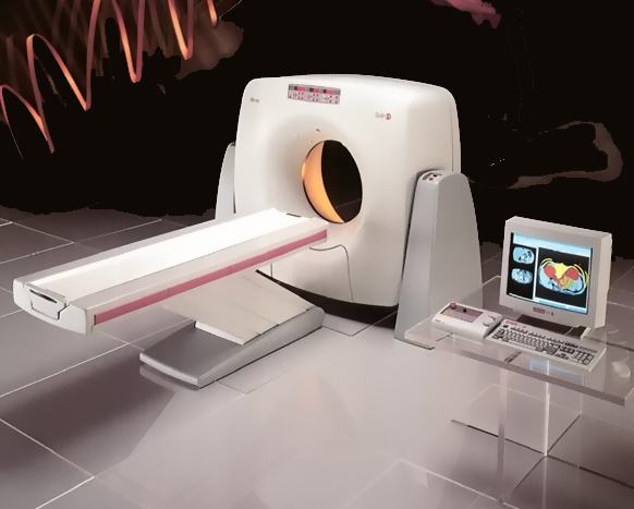
| + | http://www.radiology-equipment.com/instrumentpics/elscint.jpg | ||
Revision as of 12:52, 22 March 2008
Contents |
Computed Tomography:
When
The very first step of the long process which scientists have taken to arrive at a CT scan starts with Alessandro Vallebona in 1930. He suggested taking a single slice of the body on radiographic film, which is known as tomography. The original prototype of a CT scanner was developed in 1971 and the first brain scan was performed in England at the Atkinson Morley Hospital in 1972.
How
This is a medical method used to scan the inside of an object using a large series of two-dimensional x-rays taken around a single axis.
Use
The CT scan is an important tool used to supplement the use of x-rays and ultrasounds. It is used to create detailed images of the inside of an individual�s body including their chest, abdomen, head, and other various parts of the body. These images can then be displayed on a computer using 3-d rendering techniques.
With the use of CT scans, doctors can determine a number of factors of a patient including brain activity, bone density and many others.