Dermatology - Bright Red Rashes
From Iusmicm
Contents |
Bright Red Rashes
- There are five primary players in bright red rashes:
- Urticaria
- Erythema multiforme
- Morbilliform eruptions
- Lupus erythematosus
- Dermatomyositis
General tendencies
- In general, vascular reactions to insult can be described by color, topography, and time course:
- Color: redness = erythema, results from excessive blood flow
- Topography: raised lesions result from edmea
- Time course: a reaction can be described as dynamic = evanescent
- Note that evanescent means "soon passing out of sight or memory"
- These three classic vascular reactions (erythema, edema, and evanescent) best describe urticaria, erythema multiforme, and morbilliform reactions
Urticaria
- Urticaria is an allergic reaction that results in a bright red rash.
- Urticaria is colloquially known as "hives"
- The offending antigen is usually from a virus, a food, or a drug
- Physical factors can also induce urticaria (think "SPACE"): sun exposure, pressure, aquagenic, cold, exercise
- Pathogenesis of urticaria:
- Induction of immune response results in a dilated (erythema), leaky (edema) vascular state that can be readily changed (evanescent)
- Urticaria's EEE state is primarily driven by histamine acting on small dermal vessels
- Purpura can result if immune complexes damage endothelium to the extent that RBCs escape into the tissue
- Purpura generally lasts more than 24 hours at a single location
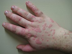
- The rash of urticaria is often described as being annular.
- Hives come and go very quickly
- Hives can be annular
- Tricyclics can be used in chronic urticaria when it is affecting the pt's mood


Erythema Multiforme
- Erythema multiforme is an allergic reaction that results in a bright red rash.
- Note that both urticaria and EM are allergic reactions.
- Erythema multiforme is distinct from urticaria by its immune complexes and its apoptotic cytotoxic T cells.
- Erythema multiforme is classified as minor and major based on how severe the manifestation.
- EM minor is usually caused by herpes simplex virus, especially when EM is recurrent.
- EM major is usually caused by drugs
- EM Major has several named causes / syndromes: Stevens-Johnson syndrome and Toxic Epidermal Necrolysis
- Both SJS and TEN are (usually) allergic reactions to drugs that result in necrolysis of keratinocytes of the lower epidermis.
- Some consider TEN a more severe form or SJS.
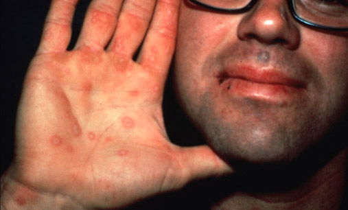
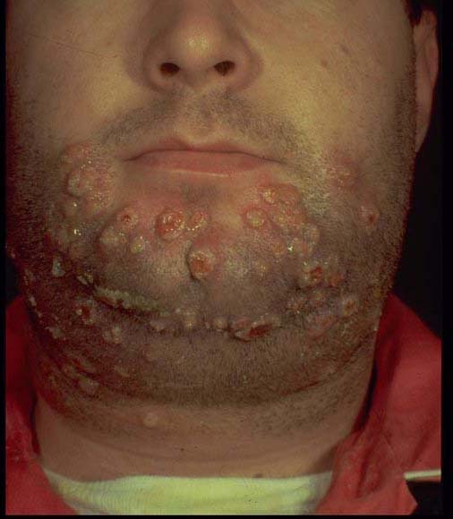
- EM presentation:
- Target lesions on palms
- Oral mucosal involvement

- http://im.unboundmedicine.com/medicine/ub/image?na=app:16010:ch18_8.bmp


- Stevens-Johnson Syndrome images:
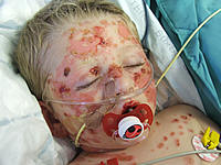
- SJS presents with widespread "targetoid" lesions
- Can have severe ocular involvement: sclerosis of cornea
- It's like a burn, so we're worried about sepsis and Na-hydration issues.
- Note that these targetoid lesions are different than the target lesions in classical EM presentation


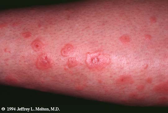
Target lesions versus Targetoid lesions
- Target lesions:
- Have concentric circles, especially when there are three concentric circles
- Have central duskiness or a central blister
- Have a relatively wide pale circle in between
- Have an outer, thin, moderately erythematous circle


- Targetoid lesions:
- Suggest zonality but don't have 3 distinct concentric circles
- In general, targetoid lesions are more serious than target lesions.


Morbilliform Reactions
- Morbilliform reaction is through to be a T-cell mediated hypersensitivity to drugs or pathogens.
- Etiology can be drugs or pathogens
- Many different drugs (including antibiotics) can cause morbilliform reactions
- Many pathogens can cause morbilliform reactions; usually enterovirus or group A streptococcus
- One diagnosis the etiology by culturing the "company" kept by the rash
- Morbilliform reaction manifests same symptoms regardless of causative agent
- Morbilliform reaction is less evanescent than multifomre erythema and urticaria
- Morbilliform reaction is characterized by truncal predominance
- Recall that urticaria could show up anywhere and multiform erythema usually involved the oral mucosa and palms of the hands
- Morbilliform is marked by its maculopapular rash


Viral Exanthems
- Most viral exanthems manifest as morbilliform reactions (except for varicella).
- Recall that an exanthem is a skin eruption secondary to systemic disease.
- Recall that varicella is a viral infection with herpes.
- Recall the viral agents that cause the classic pediatric exanthems:
- These all cause MORBILLIFORM reactions
- Rubeola (Measles, paramyxovirus infection) (four Cs): cough, coryza (watery eyes), conjunctivitis, koplik spots
- Rubeola is a systemic infection by paramyxovirus of the genus Morbillivirus
- Rubella (German Measles, rubella virus infection): 3 day progression, progresses toward cephalocaudal poles
- Roseola (Roseola Infantum, roseola virus infection): fever, followed with rash when "defervesce"
- Mononucleosis (3 Fs): fever, fatigue, (f)pharyngitis, adenopathy, liver / spleen involvement
- Kissing disease
- Fifth disease (Erythema infectiosum): slapped cheeks, fishnet erythema, recurrence
- An example of "multiple stages" seen in many viral exanthems
- Viral exanthums are characterized by: "dew drop on rose petal" formation, umbilicated vesicles, and multiple stages:


Lupus Erythematous
- Lupus erythematous is an autoimmine disease
- There are three types of lupus: systemic (SLE), discoid (DLE), and subacute cutaneous (SCLE)
- Systemic and subacute cutaneous can be induced by drugs
- All three types of SLE are considered idiopathic.
- A genetic predisposition has been demonstrated.
- SLE (systemic lupus erythematous) requires 4 of 11 ARA criteria be met.
- DLE (discoid lupus erythematous) is primarily cutaneous in manifestation.
- SCLE (subacute cutaneous lupus erythematous); subacute means it is not quite acute or chronic.
- Pathogenesis of lupus eyrthmatous is though to be UV radiation induced and involves formation of immune complexes'.
Systemic lupus erythematous
- Systemic lupus erythematous has 11 diagnostic criteria that can be divided into skin and mucus, systemic, and laboratory findings.
- To diagnose SLE, you must "DUMP the SAC on the RAC"
- Skin and mucosal criteria (DUMP):
- Discoid lupus
- Ulcers (oropharyngela, usually painless)
- Malar rash
- Photosensitivity
- Systemic criteria (SAC):
- Serositis (pleural, pericardial): inflammation of the serous tissues--things that line the lungs / heart (pleura, pericardium)
- Arthritis
- CNS (seizures, psychosis)
- Laboratory criteria (RAC)
- Renal dysfunction (Urinary analysis, elevated 24-hour urine protein levels)
- Antinuclear antibodies (or others like anti-dsDNA or anti-Smith)
- CBC (Hematology)
- DUMP the SAC on the RAC (skin / mucosa, systemic, lab)
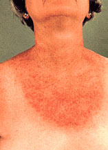
- Systemic lupus erythematous presents with a malar rash and photosensitivity
- There is sometimes "spillover" to unexposed sites, too, with the photosensitivity.
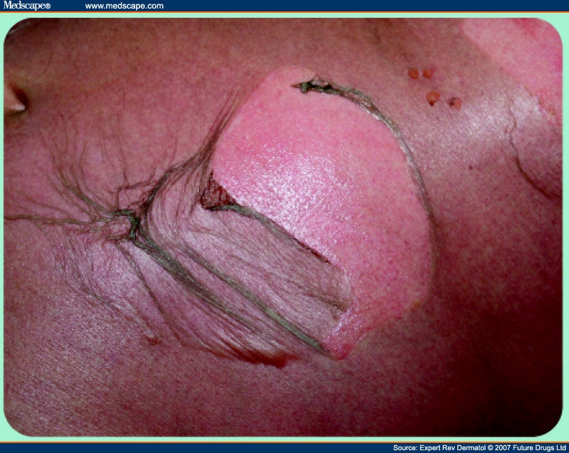
- SLE can manifest scarring alopecia
Dermatomyositis
- Dermatomyositis is an inflammation of the skin and muscle.
- Note that polymyositis is inflammation of the muscle with neurological signs but with no skin features.
- Like lupus erythematous, most dermatomyositis cases are idiopathic.
- A small subset of dermatomyositis are drug-induced.
- There is a definite role of UV radition in inducing dermatomyositis
- Dermatomyositis is characterized by involvement of the complement systems membrane attack complex (MAC)
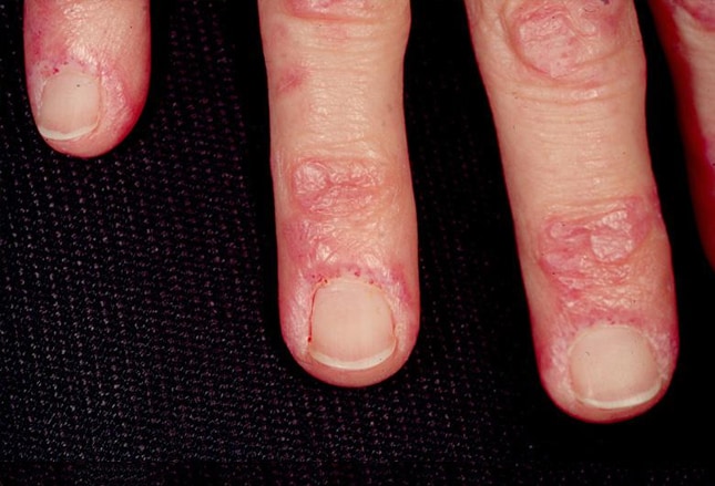
- Note that dermatomyocytis is on the IP joints of the fingers but lupus is NOT on the knuckles
- Diagnostic criteria for dermatomyositis:
- Not all 5 are required
- Skin features (to differentiate it from polymyositis)
- Proximal muscle weakness which is usually painless
- Muscle enzyme abnormalities (identified with CPK and aldolase levels)
- CPK: creatine phosphokinase; adds kinase to creatinin; MM isoform in skeletal muscle, MB isoform in cardiac muscle, BB in smooth muscle
- EMG abnormalities
- Muscle biopsy
- (currently we use MRI to dx, too)
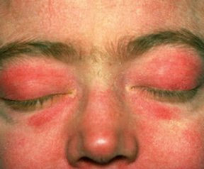
- Dermatomyositis presentation:
- Heliotrope rash (upper eyelids)
- Photosensitivity with spillover
- Poikiloderma: see next section
- Gottron's papules (area between knuckles is spared)
- Periungual telangiectasias
- Heliotrope rash (upper eyelids):
- Photosensitivity with spillover:
- Poikiloderma: see next section
- Gottron's papules (area between knuckles is spared):
- Periungual telangiectasias:


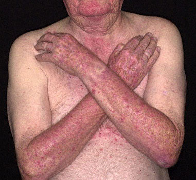


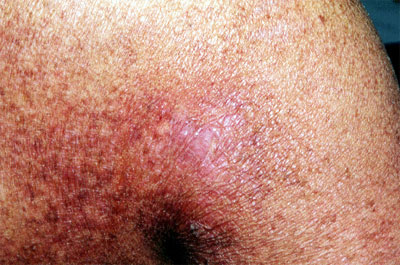
Poikiloderma
- Poikiloderma is characterized by hyperpigmentation, hypopigmentation, telangiectasias, and / or atrophy.
- Poikiloderma is seen in conjunction with dermatomyositis, UV radiation damage, and ionizing radiation damage.


Drug reactions
- Drug reactions can generate every possible different cutaneous pattern
- Rarely does clinical presentation afford enough information to dx drug-induced reaction
- Usually requires additional information like lab values
- Some patterns caused by drug reactions are considered higher risk
- These patterns have significant morbidity or have mortality associated with them
- High risk patterns include: urticaria / anaphylaxis, erythema multiforme major (SJS, TEN), morbilliform reactions (with hypersensitive syndrome characteristics), and vasculitis
- All the rest of the patterns are relatively low risk for morbidity and mortality.
An algorithm for diagnosing drug-induced cutaneous reactions
- The process is, in general, a series of controlled challenges to the pt with the drug: challenge, dechallenge, rechallenge, exlusion
- First we challenge with the drug
- An important aspect to the challenge is the time of drug administration
- This should be informed by the literature and one's own experience with the drug
- Then we "dechallenge"
- We watch for cessation of the drug reaction
- Dechallenging is options, unless the drug is essential for the pt's life
- Dechallenging will not result in cessation of recation if the reaction is irreversible
- Next we rechallenge
- This repetition gives the highest level of certainty that the reaction is drug-induced
- Rechallenging is optional
- Rechallenge is not an option if the challenge resulted in a higher-risk reaction pattern (which would advise no rechallenge for safety reasons)
- Rechallenge is not an options if there is an alternative therapy to the drug that causes a reaction (because we shouldn't give a drug we know causes an adverse reaction is there is still another potentially non-reactive drug available)
- Finally we exclude
- If the dechallenge does not generate cessation or if the rechallenge does not cause the same reaction the we exclude drug-induced reactions from the ddx.
