Exam 1 Physical Diagnosis Objectives
From Iusmicm
Revision as of 21:42, 28 February 2013 by 91.201.64.7 (Talk)
- Note, much borrowed from generous, previous IUSM medical students.
afizPK Enjoyed every bit of your post.Really looking forward to read more.
5Astck Great blog.Really looking forward to read more. Really Cool.
sR9V4Z Major thanks for the blog article.Thanks Again. Really Great.
YbuRcM Thanks so much for the blog.Much thanks again. Great.
S9qz2p Very neat blog.Thanks Again. Much obliged.
Contents |
Chapter 10
- Describe the actions and innervations of the eye and extraocular muscles.
- Identify the major symptoms of eye disease.
- Interpret the symptoms of the major diseases of the eye and apply them clinically to a patient: loss of vision, eye pain, diplopia, tearing or dryness, discharge, and redness.
- Apply the components of the physical examination of the eye to a patient.
- Clinically correlate the symptoms and physical exam findings pertaining to the eye for the following disease processes: visual field defect, red eye (acute conjunctivitis, acute iritis, narrow-angle glaucoma, and corneal abrasion), diabetes, hypertension, and papilledema.
- Give a differential diagnosis based on symptoms and/or physical exam findings pertaining to the eye.
Chapter 11
Ear
- Describe the structure and innervations of the ear.
- External ear consists of pinna and external auditory canal
- The middle ear or tympanic cavity consists of connections to the mastoid antrum and nasopharyx through the Eustachian tube
- The tympanic membrane forms the lateral boundary of the middle ear, while the cochlea forms the medial boundary
- Sound is conducted from the tympanic membrane to the inner ear by the malleus, incus and stapes
- The tensor tympani [CN V] and the stapedius [CN VII] are also located in the middle ear
- The inner ear is the end-organ for hearing and equilibrium containing the semicircular canals, vestibule and the cochlea
- The hair cells of the organ of corti convert mechanical force into electrochemical signal
- Identify the major symptoms of ear disease and how these symptoms can identify diseases involving the ear: hearing loss, vertigo, tinnitus, otorrhea, otalgia, and itching.
- Hearing loss
- conductive: blocks transmission of sound waves in external canal or middle ear; caused by wax, effusions, foreign bodies
- sensorineural: inner ear structures, auditory nerve or brain stem are dibilitated; caused by prenatal rubella, and lots of other causes.
- Patients with sensorineural talk loudly, conductive speak softly.
- Vertigo: spinning or turning in position; can be otologic, neurologic, psychological, or iatrogenic.
- Meniere’s disease: severe paroxysmal vertigo as a result of labyrinthine lesions
- Tinnitus: sensation of hearing sound such as buzzing or ringing
- Otorrhea: bloody discharge associated with carcinoma or trauma
- Otalgia: pain related to inflammatory conditions or may be referred
- Itching: from primary disorder of external ear or discharge from middle ear
- Hearing loss
- Apply the components of the physical exam of the ear to a patient.
- Inspect external ear structures: tophi, “cauliflower ear” from trauma, cancer, discharge
- Palpate the external ear structures
- Auditory acuity testing: occlude one ear and speak softly into the other; use of 512Hz fork
- Rinne test: air conduction vs bone conduction using fork; placing stem on mastoid process compared against vibratory hearing; usually AC > BC [Rhine positive]
- Weber test: compares bone conduction in both ears by placing fork in middle of forehead; sound heard on ipsi side of conductive problem and contra side of a sensorineural problem
- Example: Rinne positive on both sides -> sensorineural problem; Rinne negative on one side identifies conduction problem on that side
- Otoscopic examination: pull posterior and superior and look at the canal and tympanic membrane (should be pearly gray)
- Injection: dilation of tympanic vessels
- Retraction pocket can indicate Eustachian tube blockage and decreased pressure
- Determination of mobility of tympanic membrane: also uses otoscope; useful for middle ear infection possibility (decreased movement will be detected if fluid present)
- Clinically correlate the symptoms and physical exam findings pertaining to the ear for the following disease processes: conductive and sensorineural hearing loss, otitis media, serous otitis media, otitis externa, and vertigo. Interpret the symptoms of diseases of the ear and apply them clinically to patient.
- Acute otitis externa: p. aeruginosa
- Severe pain accentuated by manipulation of the pinna; lymphadenopathy; fever; normal tympanic membrane
- Summer time
- Bullous myringitis: localized form of external otitis
- Acute viral URI; severe pain; lesions on the skin of the deep external ear canal;
- Self-limited
- Acute otitis media: bacterial infection of the middle ear
- Usually in children;
- Pain, malaise, no pain on external manipulation, fiery red tympanic membrane;
- Winter
- Advanced acute otitis media: rupture of membrane
- Perforations can be central or marginal (more serious and may dispose to a cholesteatoma)
- May result from either otitis media or trauma
- Serous otits media:
- Adults with viral URIs or sudden atmospheric pressure changes
- Tympanic membrane appears yellowish-orange as a result of the amber-colored fluid
- Partial obstruction of the Eustachian tubes:
- Air bubbles or an air-fluid level in the middle ear
- Chronic otitis media: rupture and recurrent middle ear infections
- Foul-smelling, not usually painful
- Retraction pockets: chronic negative pressure within middle ear
- May progress to an acquired cholesteatoma
- Treat with T-tube (tympanostomy)
- Deafness:
- Child: conductive via cerumen, chronic/acute otitis media; sensorinueral via mumps, rubella
- Adult: conductive via tube blockage, viral myringitis, otosclerosis; sensorineural via Meniere’s, ototoxic drugs, acoustic neuroma
- Acute otitis externa: p. aeruginosa
- Generate a diagnosis and/or differential diagnosis based on symptoms and/or physical exam findings of the ear.
Nose
- Describe the structure of the nose.
- Illustrate how the major symptoms of nose diseases are used to identify nose diseases.
- Interpret the symptoms related to the nose and apply them clinically to a patient.
- Apply the components of the physical exam of the nose to a patient.
- Clinically correlate the symptoms and physical exam findings pertaining to the nose for the following disease processes: allergic rhinitis, sinusitis, and nonallergic rhinitis.
- Generate a diagnosis and/or differential diagnosis based on symptoms and/or physical exam findings of the nose.
Chapter 12
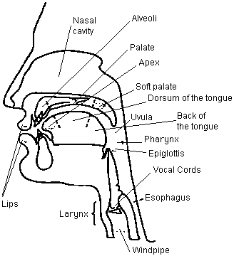
- Know the structures of the oral cavity and pharynx.
- Know the functions of the pharynx.
- Subdivisions: nasopharynx, oropharynx, hypopharynx.
- Fxn: provides swallowing, speech, and an airway.
- Know the important symptoms of disease of the oral cavity.
- Ulceration, bleeding, mass, halitosis, xerostomia (dry mouth).
- Apply the components of the physical exam of the oral cavity and pharynx to a patient.
- See cd.
- Clinically correlate the signs and symptoms of the following conditions:
- Aphthous ulcer
- Single canker sore. Most common acute oral ulcer.
- Relatively superficial w/ raised borders. On buccal or labial mucosa.
- Herpetic ulcer
- acute multiple ulcers, associated w/ vesicles.
- On mucocutaneous junction, hard palate, or gingivae.
- Crusting when bullae break.
- Chancre
- Painless, single lesion on lips or tongue.
- Lesion w/o central necrotic material.
- May have tender lymphadenitis.
- Squamous cell carcinoma:
- Single indurated sore on lips, tongue, mouth floor, or tongue (esp. on lateral borders)
- Erythroplakia of mouth floor and soft palate.
- Raised border, absence of necrotic material in crater.
- May have painless lymphadenopathy in neck.
- Candidiasis
- Burning tongue, inside of cheek or throat.
- Whitish pseudomembrane.
- Peeled off to reveal raw, red area that may bleed.
- Erythroplakia
- Painless, red area.
- Granular, red papules that bleed.
- Leukoplakia
- Painless, white area.
- Hyperkeratinized. Can’t be scraped off.
- Looks like flaking white paint. Often speckled w/ red spots.
- If associated with adenopathy, could be malignancy.
- Lipoma
- Painless mass on inner surface of cheek or tongue.
- Yellowish, soft, freely mobile.
- Lichen planus
- Usually no symptoms.
- Erosive form causes burning sores on inner cheeks and tongue.
- White reticulated papules bilaterally in lace-like pattern.
- Erosive form is hemorrhagic, ulcerated w/ possible white areas or bullae.
- May have pseudomembrane covering.
- Mucocele
- Intermittent painless swelling of lower lip, or inside cheek.
- Slightly bluish.
- Dome-shaped, freely-mobile cystic lesion.
- Hairy Tongue
- Gagging sensation.
- Large brown or black painless lesion on top of tongue.
- Elongation of filiform papillae and color change.
- Aphthous ulcer
Chapter 13
- Describe the topographical landmarks of the chest and utilize that knowledge to describe physical findings of the chest.
- Recognize the main symptoms of pulmonary disease and how these symptoms can identify disease.
- Interpret the symptoms of pulmonary disease and apply them clinically to a patient.
- Apply the components of the physical exam of the chest to a patient.
- Clinically correlate the symptoms and physical exam findings pertaining to the chest:
- Pulmonary Edema
- Pneumothorax
- Asthma
- Pneumonia
- Emphysema
- Pulmonary Embolism
- Pleural Effusion
- Generate a diagnosis and/or differential diagnosis based on symptoms and/or physical exam findings of the chest.
Chapter 17
- Describe the topographical landmarks of the abdomen and utilize that knowledge to describe physical findings of the abdomen.
- Recognize where abdominal structures are located by topographical quadrants of the abdomen.
- Recognize the main symptoms of abdominal disease and how these symptoms can identify disease.
- Interpret the symptoms of abdominal disease and apply them clinically to a patient.
- Apply the components of the physical examination of the abdomen to a patient.
- Clinically correlate the symptoms and physical exam findings pertaining to the abdomen.
- Generate a diagnosis and/or differential diagnosis based on symptoms and/or physical exam findings.


