Eye
From Iusmhistology
- started here on 04/11/11.
- NBME:
- 1/4 of the 100 questions are images.
- No questions on the eye or ear.
Contents |
[edit] The eye
[edit] Optical anatomy
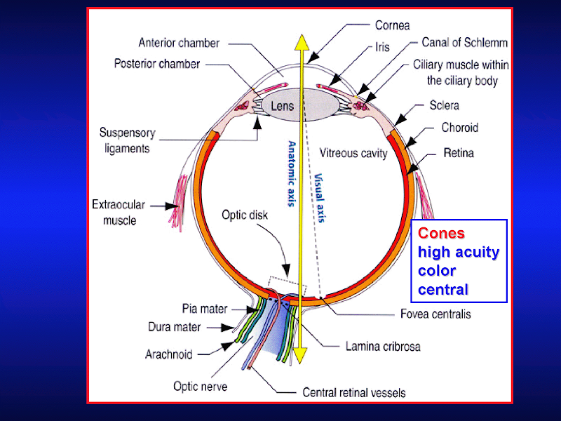
- Important anatomical features of the eyeball include:
- cornea with limbus (outer rim that holds stem cells)
- anterior chamber
- pupil
- iris
- posterior chamber
- ciliary body
- lens (with ciliary processes and zonules)
- vitreous body
- retina
- ora serrata
- fovea
- optical disc

[edit] Wall of the eye
- The wall of the eye is comprised of three layers: fibrous tunic, uvea, retina (where the retina is the inner-most layer).
- The fibrous tunic generates the cornea, the sclera, and provides continuity with the conjunctiva.
- The conjunctiva is a clear mucous membrane that lines the exterior of the sclera and cornea and is continuous along the poster aspect of the eyelid.
- The uvea is the vascular tunic (tunica vasculosa) that carries the vasculature of the eye.
- The uvea also makes the iris, ciliary bodies, and the choroid.
- The choroid is highly vascularized.
- The retina provides the sensory material of the eye.
- The retina includes the pigmented epithelium, the neural photoreceptor cells, and the neuronal integreative circuitry and supporting cells.
- Recall that photoreceptor cells come in rods and cones.
- Note that the pigmented epithelium is deep to the photoreceptors.

[edit] Eyeball function by component

[edit] Cornea
- The cornea is the most superficial layer of the eye and is exposed to the environment.
- Recall that the cornea arises from the fibrous tunic of the optical tissue.
- The cornea is transparent, avascular, and bends the incoming light to hit the lens.
- In fact the cornea bends the light (refracts) even more than the lens.
- Photorefractive keratectomy and LASIK (laser in situ keratomileusis) are two surgeries that reshape the deeper (non-epithelial) layers of the cornea to correct poor refraction that results in poor vision.
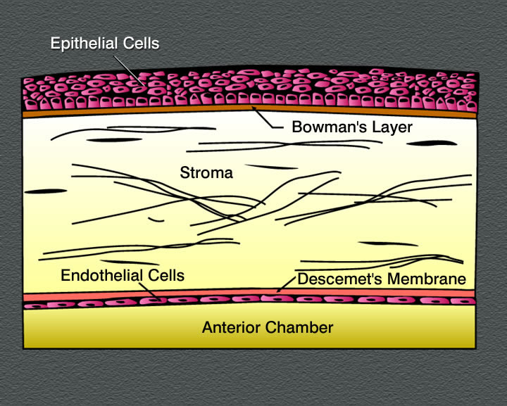
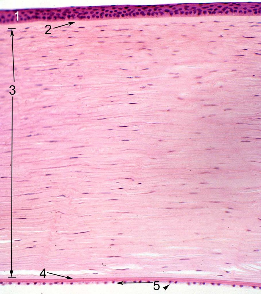
- The cornea has three cellular layers and two atypical basement membranes.
- The layers (superficial to deep): epidermis, Bowman's membrane, stroma, Descemet's membrane, and endothelium.
- With age, Descemet's membrane decreases in transparency and leads to decreased light transmission.


[edit] Epithelium of the cornea
- The epithelium of the cornea is made of stratified, squamous, non-keratinized epithelium.
- The epithelium layer is highly proliferative and has abundant pain fibers.
- The epithelial layer maintains a population of stem cells in the limbus area (the junction of the cornea with the sclera).
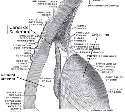
- Note that aqueous humor (from the anterior chamber) drains through the corneoscleral junction (the limbus).
- We call the corneoslceral junction (the limbus) a trabecular meshwork and the site of Schlemm's canal.


[edit] Stroma of the cornea
- The stroma is also called the substantia propria.
- There are 50-60 layers of cells making the stroma the most substr
- The stroma is series of layers of fibrocytes, proteoglycans, and ECM fibers with alternately oriented collagen fibers.
- Note that collagen of the stroma is of type 1 and 5 and are non-fibrillar.

[edit] Endothelium of the cornea
- The endothelium of the cornea contains leaky intercellular junctions that allow exchange of fluid with the anterior chamber.
- Drainage by the endothelium keeps the stroma relatively dry.
- Note that fluid transport by the endothelium contributes to the transparency of the cornea.


[edit] Uvea
- Recall that the uvea is one of the three layers of the eye: the tunica fibrous, the uvea, and the retina.
- Recall that the uvea is also called the tunica vasculosa and carriers the vasculature of the eye.
- The uvea includes the iris, the ciliary bodies, and the choroid.
- Just as the cornea is the anterior portion of the fibrous tunic and is completed posteriorly by the sclera, the iris and ciliary body are the anterior aspect of the tunica vasculosa and are completed posteriorly by the choroid.

[edit] Choroid of the uvea
- Recall that the choroid is the posterio-lateral aspect of the uvea (tunica vasculosa).
- The choroid carries vasculature to the retina and the sclera.
- The choroid is highly pigmented and found between the sclera (part of the tunica fibrous) and the retina.
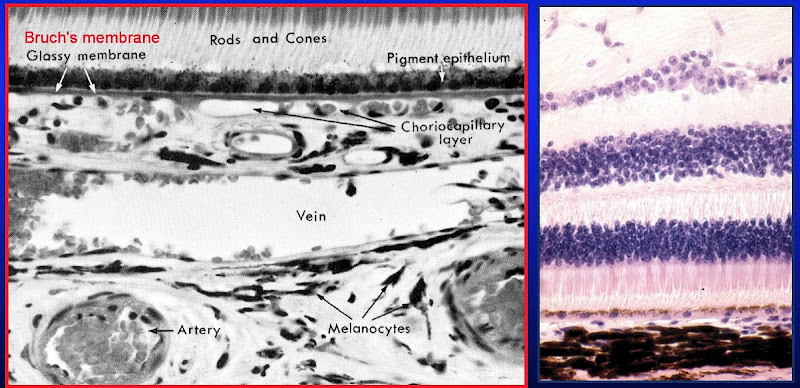
- The choroid has three layers: sclera vasculature, retinal vasculature, and Brunch's membrane.
- The retinal vasculature layer is also called the choriocapillary layer.
- Note that these layers are indistinguishable in the lab.
- The choriod layer is separated from the retina by the Bruch's membrane; that is, Brunch's membrane is the deepest aspect of the choroid part of the tunic vasculosa (uvea).
- Bruch's membrane is also sometimes called a glassy membrane.

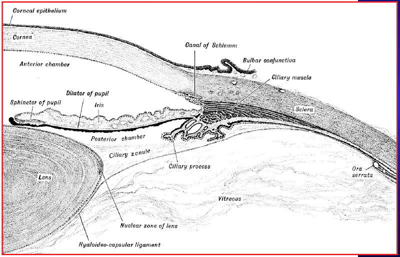
[edit] Ciliary body
- The ciliary body is a thickening of the vascular layer near the anterior aspect, radially from the lens.
- Ciliary bodies have two structures: ciliary zonules (ligaments) and ciliary processes.
- Ciliary zonules (suspensory ligaments):
- Ciliary ligaments project from the tunica vasculosa to the lens capsule and control the flattness / bulging of the lens in order to focus light (a process called accomodation.
- Upon contraction of the ciliary muscle, the many ciliary zonules are brought closer together and anterior which actually allows the lens to relax.
- Think of the ciliary muscle as a sphincter and the relationship with zonules and the lens makes sense.
- Far vision requires a flattened lens (think flat like a frisbee which you hope will go "far") so the ciliary muscle relaxes, the zonules tense, and the lens is pulled into a flatter shape.
- Near vision requires a bulged lens so the ciliary muscle contracts, the zonules relax, and the lens relaxes into a bulge.
- Recall too that near vision gives eye strain which is the excessive contraction of the ciliary muscles.
- Oxytalin fibers are used to attach the basement membrane of the non-pigmented epithelium of the ciliary body (part of the uvea) to the basement membrane (called the lens capsule) of the lens.

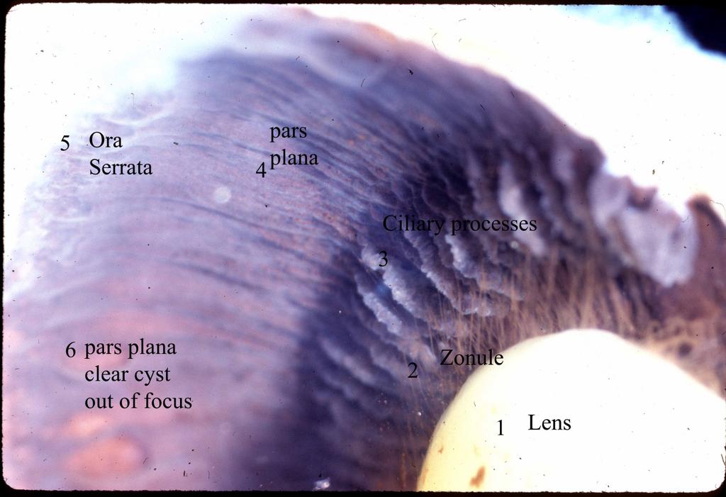
- Ciliary processes:
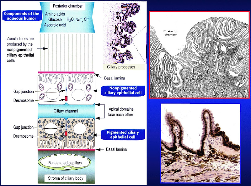
- Ciliary processes primarily function to generate aqueous solution.
- Aqueous solution provides nutrition to the lens and maintains intraocular pressure.
- Aqueous solution has little protein, some glucose, and similar ion concentration as plasma.
- Ciliary processes are an extension of the tunica vasculosa (the uvea) and are highly vascularized.
- There are two layers to the ciliary process tissue: deep layer (pigmented) and surface layer (secretory, non-pigmented).
- The pigmented layer (deep layer) of the ciliary process is continuous with the pigmented layer of the retina.
- The non-pigmented layer's (secretory, surface layer) apical surface faces the pigmented layer and the basolateral surface has lots of folds and borders the posterior chamber.
- We call the space between the pigmented and non-pigmented cells the ciliary channel and consider it a potential space.
- Blood-aqueous barrier: occluding junctions at the apex of the ciliary process's surface epithelium keeps blood and aqueous solution from mixing.
- Note that glaucoma results from blocked drainage of aqueous fluid and the resulting elevation of intraoccular pressure.
- Ciliary processes primarily function to generate aqueous solution.

.jpg)
[edit] Iris
- The iris the most anterior aspect of the uvea (the vascular layer).
- Recall that the uvea is the middle layer of the three layers of the eyeball: tunica fibrous, tunica vasulosa (uvea), and the retina.
- The iris divides the eyeball into two chambers: the anterior and posterior chambers.
- The iris's job is to regulate the amount of light entering the eye ball.
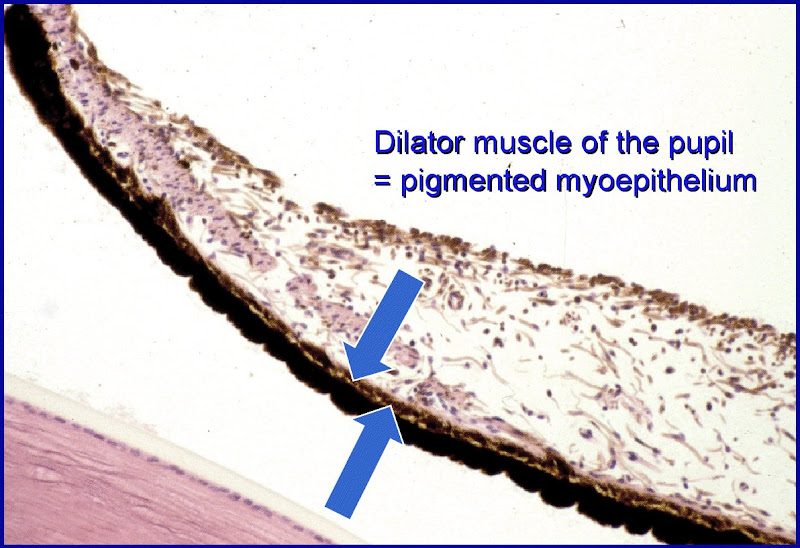
- The posterior aspect of the iris has epithelial and myoepithelial layers that provide the pigment of eye color and the movement of the iris, respectively.
- Note that the posterior epithelium is highly pigmented but does not determine eye color.
- Note that the posterior epithelium is one and the same as the myoepithelium: it has muscle fibers in it and is responsible for dilating the pupil.
- The iris is primarily composed of stroma with an epithelial covering only on the posterior surface.
- The stroma is highly vascular (recall that the iris is part of the tunica vasculosa) and has fibroblasts, connective tissue, melanocytes, and myocytes.
- The malanocytes of the stroma determine eye color.
- The stroma is responsible for constricting the pupil.
- Note that the stroma contains the constrictor muscle of the pupil and the myoepithelium contains the dilator muscle of the pupil.
- Note that dilation is sympathetic and contraction is parasympathetic.
[edit] Eye color
- Eye color is determined by the quantity and distribution of pigment cells in the stroma and posterior epithelilum of the iris.
- Pink: complete lack of pigment; albinism (lack of tyrosinase or a defect in melanosome transport, RAf27a)
- Blue < grey / green < brown => less to more pigment.
- Heterochromia iridium is the presence of distinct regions of different color in the iris.

[edit] Lens
- The lens of the eye is biconcave, transparent, and avascular.
- The lens capsule is the basement membrane of the lens and sits on the outer surface.
- Note that the capsule is composed of both lens fiber cells and epithelial cells.
- The lens has an epithelium but only on the anterior surface just as the iris only has an epithelium but only on the posterior surface.
- The epithelium on the anterior side of the lens is simple cuboidal.
- Note that the lens epithelium does not have occluding junctions.
- The stroma of the lens is composed of highly specialized cells (called lens fibers) that span the anterior-posterior axis and refract light well because of their high concentration of crystallin a cellular protein.
- The cells at the center of the lens contain the highest concentration of crystallin.
- These stromal cells are 7-10 mm in length.

- In the case of cataracts, lens fibers (the cells that span ant-post and have crystallin) turn opaque and refract light poorly.
- In the case of presbyopia, the lens loses elasticity (some loss is normal during aging) such that the pt cannot accomadate well and thus has poor near vision.
[edit] Retina
- The retina is the inner-most of the three layers of the eye (tunic fibrous, tunica vasculosa, and retina).
- The retina has two functional layers: the pigmented epithelium on the outside (next to Brunch's membrane from the tunic vasculosa = uvea) and the neural retina.
- Note that the neural retina and the pigmented epithelium are not well connected and thus retinal detachment often occurs between these two layers.
- The epithelium is essentially a support tissue for the neural retina.
- The unlike the tunica fibrous and (almost) the tunica vasculosa, the retina does not continue all the way anteriorly.
- The anterior apex of the retina is called the ora serrata.
[edit] Pigmented epithelium of the Retina
- The pigmented epithelium of the retina is simple cuboidal and is adjacent to the inner-most layer of the uvea (Bruch's membrane).
- Recall that Bruch's membrane is also sometimes called a glassy membrane.
- The pigmented epithelium serves to support the neural retina (which is deeper) and thus its apical membrane faces the neural retina:
- secretes nutrient-rich fluid onto the nueral retina
- Note that secretion is enabled via Na-K ATPase.
- produces melanin to absorb light and reduce scatter
- cleans up material shed by rods an cones (phagocytosis, discs)
- concentrates and estrifies vitamin A for easy reproduction of rhodopsin by rods
- secretes nutrient-rich fluid onto the nueral retina
[edit] Neural retina
- The neural retina is the inside (deep) layer of the retina's two layers (pigmented epithelium and neural retina).
- The cells that make up the neural retina include: photoreceptors (rods and cones), supporting cells, and integrative neurons.
- Rods and cones have their own anatomy related to their function (from closest to the field of view to closest to the brain): synaptic body, inner segment, outer segment.
- The synaptic body is in contact with bipolar cells of the retina.
- Note that the location of synapse from the photoreceptor to the bipolar cell is closer to the field of view than the location of photoreception.
- The inner segment:
- The outer segment contains the photosensitive apparatus, has highly modified cilium, and is in contact with the pigmented epithelium.
- Note that the outer segment is closes to the brain.
- The synaptic body is in contact with bipolar cells of the retina.


- Rods versus cones
- Rods are specialized for low light vision while cones are specialized for high light and color vision.
- "C" is for color.
- Rods use rhodopsin while cones have three distinct pigments for three colors: red, green, and blue.
- Rods are associated with the "peripheral" areas of the eye while the cones are found in higher density at the "center" region.
- Note that "peripheral" and "central" are quoted because we don't really mean the "center" we mean near the fovea centralis--the focal point of highest acuity.
- It makes sense that the cones are concentrated in the "central" area because they are high acquity photoreceptors and the center is where our focal point usually sits.
- The fovea is the cones-only area of the retina and the location of highest visual acquity.
- Rods are specialized for low light vision while cones are specialized for high light and color vision.
| Attribute | Rods | Cones |
|---|---|---|
| Function | Night vision (scotopic), low acquity | Day vision (photopic), color, high acquity |
| Photopigment | Rhodopsin | 3 pigments, 3 wavelength specificities |
| Distribution | "Peripheral" | "Central" |
| Population count | 120 million | 6 million |

[edit] Lab
- Note that the Bowman's membrane in the cornea is large and thick and separates the stroma from the epithelium.
- stopped here on 04/11/11.
