Skin
From Iusmhistology
(Difference between revisions)
(Created page with '*started here on 04/06/11. ==Skin== *The skin is the heaviest, largest organ of the boyd. *Skin is involved in protection, temperature regulation, hydration, excretion, and pro…') |
|||
| Line 373: | Line 373: | ||
**http://faculty.une.edu/com/abell/histo/apocrinesgw.jpg | **http://faculty.une.edu/com/abell/histo/apocrinesgw.jpg | ||
**http://instruction.cvhs.okstate.edu/histology/HistologyReference/imagesco/skinglands3F.jpg | **http://instruction.cvhs.okstate.edu/histology/HistologyReference/imagesco/skinglands3F.jpg | ||
| + | |||
| + | ===Understand the mechanism of skin repair=== | ||
| + | *Repair of a wound to the epidermis / dermis requires proliferation at both layers and the contribution of clotting. | ||
| + | *Increased vascular flow at the site of wound contributes to formation of a clot known as a scab, made of fibrin, which generates an initial scaffold for epidermal and dermal cells to follow in remodeling the area. | ||
| + | *The dermis is responsible for cleaning up the damaged cartilage (via macrophages) and proliferating new fibroblasts to generate new connective tissue. | ||
| + | *The epidermis contributes basal cells (recall that they are mitotically active) near the wound to migration and proliferation at the wound to generate new epithelial layers. | ||
| + | |||
| + | |||
| + | *When the whole epidermis and dermis are damaged, cells from the base of the hair follicles and sabaceous glands can migrate and proliferate to regenerate the dermis and epidermis. | ||
| + | **Indeed, we can even '''generate iPSCs''' from these adult stem cells. | ||
| + | |||
| + | |||
| + | http://journals.cambridge.org/fulltext_content/ERM/ERM5_08/S1462399403005817sup005.gif | ||
| + | |||
| + | ===Describe the histological findings in common skin diseases=== | ||
| + | *We will look at the histology of three classes of integument ills: blistering, psoriasis, and skin cancer. | ||
| + | |||
| + | ====Blistering==== | ||
| + | *Blistering occurs because of intercellular connection loss or because of a loss of connection at the epidermis-dermis junction. | ||
| + | *Abnormalities at the '''epidermis-dermis junction''' are called '''bullous pemphigoid'''. | ||
| + | **http://www.dermpedia.org/files/images/Bullous_pemphigoid_3.jpg | ||
| + | *Abnormalities of intercellular junctions are called '''pemphigus'''. | ||
| + | **http://library.med.utah.edu/kw/derm/mml/24820016.jpg | ||
| + | |||
| + | ====Psoraisis==== | ||
| + | *Psoriasis occurs when cells of the basal and spinosum layers demonstrate '''excessive proliferation''' and decreased cycle time which leads to '''increased thickness'''. | ||
| + | *One can identify psoriasis by the presence of '''nuclei in the stratum corneum'''; this finding is called '''parakeratosis'''. | ||
| + | **Recall that cells of the stratum corneum do not usually have nuclei. | ||
| + | **Recall that we called the tongue epithelium '''perakeratinized''' because it had significant keratinization, differential staining, and yet the nuclei remained present. | ||
| + | |||
| + | http://www.drmihm.com/pictures/ACF1E7.jpg | ||
| + | |||
| + | http://www.webpathology.com/slides/slides/ExtGenitalia_Pagets1.jpg | ||
| + | |||
| + | http://dermatology.cdlib.org/126/unknown/eyelid/2.jpg | ||
| + | |||
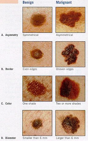
| + | ====Skin cancer==== | ||
| + | *There are three major types of skin cancer, named for the origin of the tumor cells: basal cell carcinoma, squamous cell carcinoma, and malignant melanoma. | ||
| + | |||
| + | http://www.aafp.org/afp/2004/1015/afp20041015p1481-f1.gif | ||
| + | |||
| + | =====Basal cell carcinoma===== | ||
| + | *Note the excessive proliferation of the basal cells often drives them downward into the dermis. | ||
| + | |||
| + | http://skincancer-fact.com/wp-content/uploads/2009/10/Basal_cell_carcinoma_-skin_cancer-picture.jpg | ||
| + | |||
| + | http://www.orlandoskindoc.com/Basal-cell-carcinoma-large.jpg | ||
| + | |||
| + | http://www.trihealth.com/ser/cancer/images/Basal_cell_carcinoma.jpg | ||
| + | |||
| + | http://www.healthpm.com/wp-content/uploads/2010/09/basal_cell_carcinoma.jpg | ||
| + | |||
| + | http://missinglink.ucsf.edu/lm/DermatologyGlossary/img/Dermatology%20Glossary/Glossary%20Histo%20Images/basal_cell_carcinoma_high_power.jpg | ||
| + | |||
| + | =====Squamous cell carcinoma===== | ||
| + | *Note the aberrant growth in the stratum spinosum. | ||
| + | |||
| + | http://www.medicalook.com/diseases_images/squamous_cell_carcinoma.jpg | ||
| + | |||
| + | http://missinglink.ucsf.edu/lm/DermatologyGlossary/img/Dermatology%20Glossary/Glossary%20Histo%20Images/squamous_cell_carcinoma_in_situ_high_power.jpg | ||
| + | |||
| + | =====Malignant melanoma===== | ||
| + | *Note the excessive number of melanocytes. | ||
| + | *'''Mohs surgery''' first removes the visible melanoma and then uses microscopic examination of the tumor site and a precise map and excision technique to remove the '''roots of the cancer'''. | ||
| + | |||
| + | http://www.prlog.org/10263280-malignant-melanoma.jpg | ||
| + | |||
| + | http://sunsafekids.tripod.com/sitebuildercontent/sitebuilderpictures/melanomas.jpg | ||
| + | |||
| + | http://www.nature.com/modpathol/journal/v18/n8/images/3800395f1.jpg | ||
| + | |||
| + | http://rad.usuhs.mil/derm/lecture_notes/Images/melanoma_histo.jpg | ||
| + | |||
| + | ====Griscelli syndrome==== | ||
| + | *Griscelli syndrome is also called "silver baby syndrome". | ||
| + | *Griscelli syndrome can result from a defective Rab27a protein which is part of the transport complex that moves '''melanosomes''' along '''microtubules''' for cyotocrine passage to other cells. | ||
| + | *Patients with silver baby syndrome have '''hypopigmentation''' and also '''immunodeficiency'''. | ||
| + | |||
| + | |||
| + | *stopped here on 04/06/11. | ||
Revision as of 15:30, 12 April 2011
- started here on 04/06/11.
Skin
- The skin is the heaviest, largest organ of the boyd.
- Skin is involved in protection, temperature regulation, hydration, excretion, and production of hormones, cytokines, and growth hormones.
Describe the basic histological structure of the skin
- There are 2 + 1 layers to the skin: the epidermis, the dermis, and the hypodermis.
- The epidermis originates from the ectoderm and is composed of epithelial cells.
- The dermis originates from the mesoderm.
- The hypodermis is not considered part of the skin proper and contains adipose tissue.


Identify the cell layers that constitute the epidermis
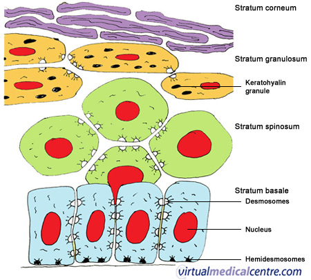
- The epidermis contains 4 layers in most locations and 5 layers on the hands and feet.
- The epidermis of the hands and feet is called thick skin and is hairless and contains a fifth layer.
- From deep to superficial, the 5 layers of thick skin are: basale, spinosum, granulosum, lucidum, and corneum (BSGLC).
- The epidermis is not vascularized so the cells must rely on diffusion of nutrients from deep to superficial.
- Therefore, it makes sense that deeper cells are more alive and the shallower cells are more dead-like.
- Cellular progression from deep to superficial is also why it makes sense to talk about the layers from deep to superficial.


Stratum basale
- The stratum basale is also called the stratum germinativum.
- This base layer of epidermis is made of a single layer of mitotically active columnar or cuboidal cells.
- Recall that many adult stem cells live on basement membranes.
- The cells of the basal layer are connected to the basement membrane via hemidesmosomes.
/Integument/Thick%20Skin%20-%20Tutorial/F%20-%20Thick%20Skin%2040X-3-Epidermal%20Layers.jpg)
Stratum spinosa
- The spinosa appears as though there are a series of "spears" aligned within.
- This spinous look results from several layers of of cells connected by desmosomes.


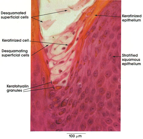
Stratum granulosmum
- The stratum granulosum contains cells with granules which usually implies they have some specific role--and they do.
- The granule-containing cells of the granulosum contain filaggrin, and intermediat filaments that help to form the tonofibrils along with keratin.
- Note that these granules are called keratohyline granules.
- The cells of the granulosum are flattened polygonal cells--found in 3 to 5 layers.
- Epidermal cells of the granulosum are generally basophilic.
- Note that the granulosum is thickened in thick skin.
- There is a second type of granule (called lamellar granules) that contain lipids that are excreted into the connective tissue to provide a barrier to water-loss.
- Just like "lamellae" in chloroblasts, lamellar granules have a sort of stacked-bags look


Stratum lucidum
- The stratum lucidum is only present in thick skin.
- The stratum lucidum contains only a few layers of very flattened eosinophilic epidermal cells.
- The lucidum (ironically) stains darkly.
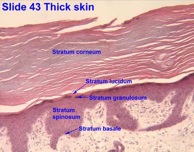
Stratum corneum
- The stratum corneum contains 15-20 layers of very thin, highly keratinized cells.
- These cells are called squames.
- Note that corneum means horny; think of a rhinocerus's horn--it is keratinized skin.
/Integument/Thick%20Skin%20-%20Tutorial/D%20-%20Thick%20Skin%2040X-1-Epidermal%20Layers.JPG)
Dermo-epidermal junction
- Recall that the stratum basal is the deepest layer of the epidermis and that the dermis is deep to the epidermis.
- Recall, too, that the epidermal cells of the basal layer are connected to the basement membrane via hemidesmosomes.
- Recall that the dermis contains lots of connective tissue.
- The dermis contains lots of type 4 collagen.
- The superficial aspect of the dermis that is attached to the basement membrane of the epidermis is the lamina densa.
- The lamina densa of the dermis and the basement membrane of the epidermis are connected via anchoring filaments and anchoring fibrils.
- Note that anchoring filaments are composed of type 7 collagen.

Describe the cellular components of the epidermis and their functions
- There are four major cells of the epidermis: keratinocytes, melanocytes, langerhan cells, and merkel cells.
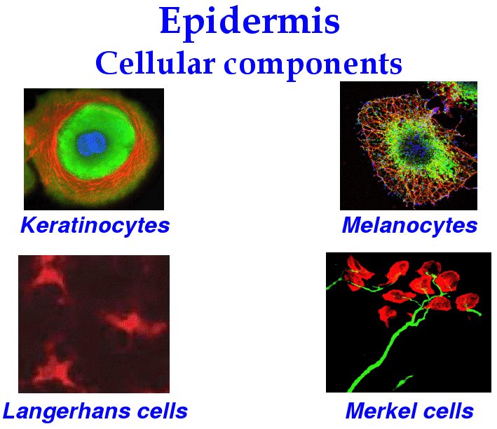
Keratinocytes
- Keratinocytes are the most numerous cell of the epidermis and primarily serve a structural role.
- Keratinocytes produce the protein keratin.
- Keratinocteys also help produce the water barrier with their tight junctions.
- Keratinocytes are found in all layers of the epidermis and take many shapes depending on their location.

Melanocytes
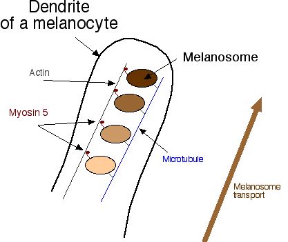
- Melanocytes reside in and near the stratum basale and have dendritic projections that weave through neighboring keratinocytes.
- Melanocytes are derived from neural crest cells and function to generate the pigment melanin which protects cells from UV damage.
- Melanocytes generate melanin and pass it to neighboring cells via cytocrine secretion upon MSH signaling.
- Note that melanin is produced from tyrosine via tyrosinase in a specialized intracellular organelle called a melanosome.
- Melanosomes are said to "mature" as they are produced; they turn from a light, circular shape to a dense cucumber shape.
- Eumalanosomes and phaeomelanosomes give color to bird and dinosaur feathers.
- Albino organisms often lack tyrosinase and thus cannot generate melanin.
- Note that melanin is produced from tyrosine via tyrosinase in a specialized intracellular organelle called a melanosome.


Langerhans cells
- Langerhans cells are derived from bone marrow and thus it makes sense that they serve an immunological role.
- Langerhans cells bind, process, and present antigens to T cells with their star shaped cell bodies.
- Langerhans cells reside primarily in the spinosum layer (think "spines and chinese are for killing bad guys!).


Merkel cells
- Merkel cells (like melanocytes) are found primarily in the basal layer, which makes sense because they are a sensation cell that needs to be near a nerve ending.
- Note that Merkel cells are found primarily in thick skin where touch needs to be highly sensitive.
- Merkel cells, along with the expanded terminal bulb of afferent, myelinated nerves, form the Merkel's corpuscle which detects touch as a mechanoreceptor.
- The Merkel cells contain dense-cored neurotransmitter granules.

Describe the structural organization of the dermis
- Recall that the dermis is a connective tissue layer (collagen 4) that connects the epidermis to the hypodermis.
- The dermis has two layers (superficial to deep): papillary layers and reticular layer.
Papillary layer of the dermis
- The papillary layer is a delicate layer of connective tissue called papillary because of the papilla it forms as it protrudes up into the epidermis.
- Connective tissue of the papillary layer is composed of type 1 collagen, type 2 collagen, and elastic fibers.
- Recall that type 7 collagen binds the dermis to the basal lamina (the basement membrane) that separates the epidermis and dermis.
- It is the papilla between the epidermis and papillary layer of the dermis that generate fingerprints.
- When you think of fingerprints think of Mark Twain and Juan Vucetich.
- The papillary layer contains nerves and vessels but they do not penetrate the epidermis.
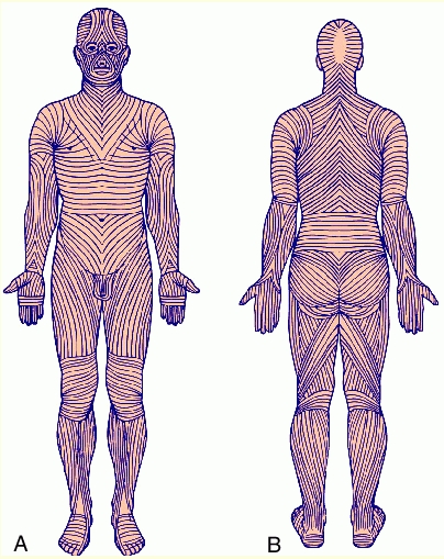
Reticular layer of the dermis
- The reticular layer of the dermis is a sturdier connective tissue than the papillary layer and is less cellular.
- The reticular layer is composed of type 1 collagen and regularly oriented elastic fibers (called Langer's lines).
- The reticular layer is always thicker than the papillary layer; though the thickness of the reticular layer can vary depending on the location.


Identify other structures in the skin
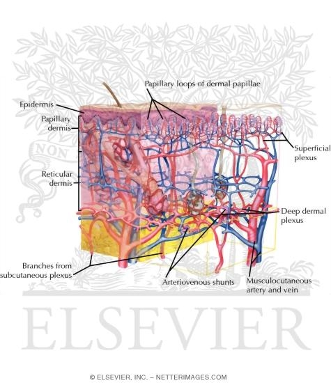
Vessels
- The vascular layout of the skin serves two functions: to provide nutrient and waste exchange and to regulate body temperature.
- There are two plexuses of arteries: between the two layers of the dermis (papillary and reticular) and between the dermis and hypodermis.
- There are three plexuses of veins: between the two layers of the dermis (papillary and reticular) and between the dermis and hypodermis (just like the arteries) and a third plexus in the middle of the dermis.
- Recall that the reticular layer is thicker than the papillary layer so even though there is a vascular plexus between the two, there is also a veinous plexus deeper in the reticular layer.
- Arteriovenous anastomoeses serve as the shunts that can be opened or closed for temperature reasons--cooling or conserving, respectively.
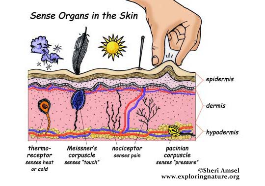
Sensory receptors
- There are four types of sensory receptors in the integumentary system (skin): free nerve endings, pacinian corpuscles, meissner's corpuscles, and ruffini's corpuscles.
Free nerve endings
- Free nerve endings are found in the stratum granulosum and detect fine touch, heat, and cold.
- Free nerve endings are not surrounded by connective tissue or schwann cells.


Pacinian corpuscles
- Pacinian corpuscles are nerve endings surrounded by an oval encapsulation of connective tissue in the deeper dermis and hypodermis.
- Pacinian corpuscles detect vibration.
- Pacinian corpuscles are myelinated.
- Think maraca shaped and all that vibration!


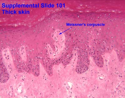
Meissner's corpuscles
- Meissner's corpuscles are found in the papillary layer of the dermis and are sensitive to low frequency stimuli.
- Doesn't "meissner" sound like an old miser with a low, grumpy voice?
- Meissner's corpuscles are shaped like tapered mitochondria and are oriented perpendicular to the skin.


Ruffini's corpuscles
- Ruffini's corpuscles are simple mechanoreceptors and have an "elongated fusiform shape".

Sensory receptor images


Hair follicles
- The hair follicle is responsible for production of hair is a structure composed of epidermal and dermal structures and cell types.
- Hair follicles originate from the invagination of epithelial cells downward to meet the papillary extension of dermal cells.
- At the hair follicle a specialized layer called the glassy membrane (a type of basement membrane that is thickened and keratinized) separates the epidermis and the dermis.
- Recall that a basement membrane separates the epidermis and dermis elsewhere, also, but that it is not keratinized.
- Note that the glassy membrane is acellular, like all basement membrane.
- During hair growth, the follicle has a bulbous end at the deepest part.
- Vasculature that arises from the dermal layer up through the dermal papilla services the bulb of the hair follicle.
- Surrounding the bulb (and originating from the epithelial layer) are melanoctyes which generate the melanin that dictates hair color.
So, why is pubic hair always dark, even if head hair is light? Do these melanocytes have more MSH receptors or some intracellular difference that generates a darker version of melanin?
- Hair has three layers (from outside in): cuticle, cortex, and medulla.
- The cuticle is the outer most and is comprised of squamous cells.
- The cortex contains cuboidal cells that differentiate into keratinized cells.
- The medulla contains large cells with vacuoles that are moderately keratinized.
- Sebaceous glands are often associated with hair follicles and produce sebum as their secretion.
- Sebum is released into the infundibulum which is a pilosebaceous canal that surrounds the base of the growing hair.


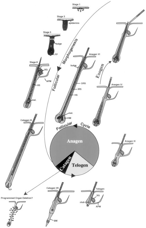
Phases of hair growth
- There are three phases of hair growth: anagen, catagen, and telogen.
- Anagen is active growth.
- Catagen is apoptosis-driven involution
- Telogen is the "resting phase" in which there is no more apoptosis but a new hair has yet to begin growing.

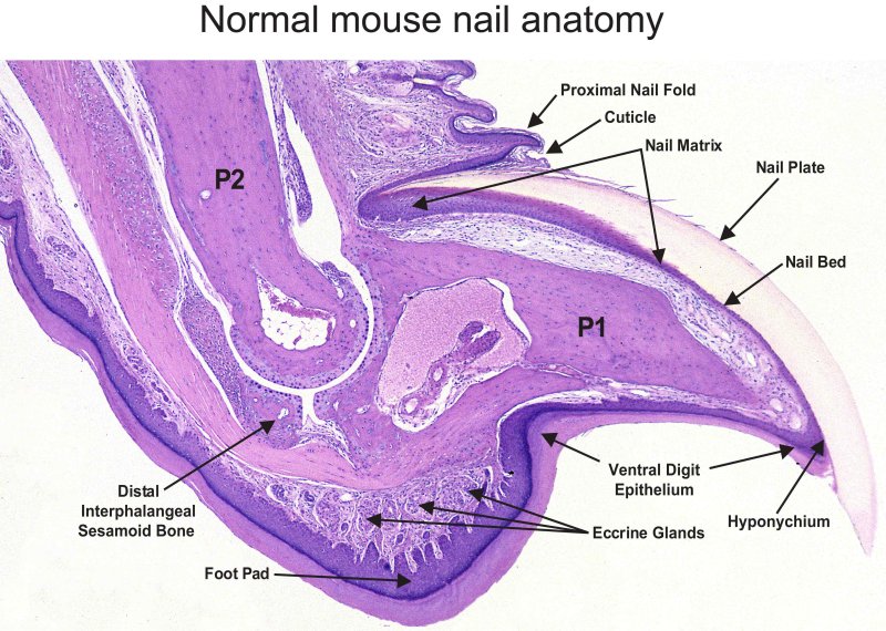
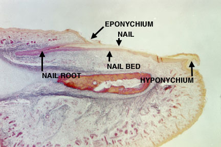
Nails
- The nail (the nail plate) is a structure of specialized stratum corneum that has hard keratin that does not desquamate like the skin corneum layer.
- The nail plate sit in the nail bed which is formed by the stratum basale and spinosum.
- Recall that the layers of the skin are normally BSGLC and here we have corneum sitting on spinosum and basal--the granulosum is missing.
- The nail bed is surrounded by the nail matrix which has a variety of cells: dividing cells that will cornify to become nail plate, melanocytes, merkel cells, langerhans cells, and stem cells.
- It makes sense that there are sensory cells in the nail matrix because sometimes you hit your nail plate just right and it hurts so bad!
- The nail root is the proximal end of the nail plate to which cornified cells are added to generate nail growth.


Glands
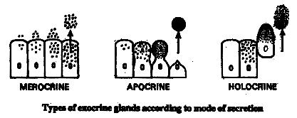
- There are two types of glands in the integument: sebaceous glands and sweat glands.
- Sebaceous glands use an holocrine method of secretion; that is, they produce more and more of their secretion and then rupture their cell membrane and spill the contents of the lumen.
- Sweat glands come in two forms: merocrine (eccrine) and apocrine.
- Merocrine (eccrine) secretion is the dumping of vesiclular content.
- Apocrine is the release of part of the cell by division of the cell membrane.

Sebaceous glands
- Sebaceous glands use holocrine secretion: the release of the contents of the lumen.
- Recall that sebaceous glands are often associated with hair follicles.


Sweat glands
- Sweat glands come in two forms: merocrine (eccrine) and apocrine.
| Attribute | Merocrine | Apocrine |
|---|---|---|
| Secretion method | Merocrine | Apocrine and merocrine |
| Distribution | Widely distributed | Axillary and perineal regions only |
| Lumen size | Small lumen | Large lumen |
| Epithelial type | Stratified cuboidal | Simple cuboidal |
| Innervation | Cholinergic fibers (ach) | Adrenergic (cats) |
- Merocrine (eccrine):
- Recall that eccrine (merocrine) secretion is the dumping of vesiclular content.
- Merocrine sweat glands are widely distributed
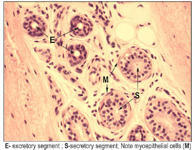

- Apocrine:
- Recall that apocrine secretion is the release of part of the cell by division of the cell membrane.


Understand the mechanism of skin repair
- Repair of a wound to the epidermis / dermis requires proliferation at both layers and the contribution of clotting.
- Increased vascular flow at the site of wound contributes to formation of a clot known as a scab, made of fibrin, which generates an initial scaffold for epidermal and dermal cells to follow in remodeling the area.
- The dermis is responsible for cleaning up the damaged cartilage (via macrophages) and proliferating new fibroblasts to generate new connective tissue.
- The epidermis contributes basal cells (recall that they are mitotically active) near the wound to migration and proliferation at the wound to generate new epithelial layers.
- When the whole epidermis and dermis are damaged, cells from the base of the hair follicles and sabaceous glands can migrate and proliferate to regenerate the dermis and epidermis.
- Indeed, we can even generate iPSCs from these adult stem cells.

Describe the histological findings in common skin diseases
- We will look at the histology of three classes of integument ills: blistering, psoriasis, and skin cancer.
Blistering
- Blistering occurs because of intercellular connection loss or because of a loss of connection at the epidermis-dermis junction.
- Abnormalities at the epidermis-dermis junction are called bullous pemphigoid.
- Abnormalities of intercellular junctions are called pemphigus.


Psoraisis
- Psoriasis occurs when cells of the basal and spinosum layers demonstrate excessive proliferation and decreased cycle time which leads to increased thickness.
- One can identify psoriasis by the presence of nuclei in the stratum corneum; this finding is called parakeratosis.
- Recall that cells of the stratum corneum do not usually have nuclei.
- Recall that we called the tongue epithelium perakeratinized because it had significant keratinization, differential staining, and yet the nuclei remained present.


Skin cancer
- There are three major types of skin cancer, named for the origin of the tumor cells: basal cell carcinoma, squamous cell carcinoma, and malignant melanoma.

Basal cell carcinoma
- Note the excessive proliferation of the basal cells often drives them downward into the dermis.


Squamous cell carcinoma
- Note the aberrant growth in the stratum spinosum.


Malignant melanoma
- Note the excessive number of melanocytes.
- Mohs surgery first removes the visible melanoma and then uses microscopic examination of the tumor site and a precise map and excision technique to remove the roots of the cancer.


Griscelli syndrome
- Griscelli syndrome is also called "silver baby syndrome".
- Griscelli syndrome can result from a defective Rab27a protein which is part of the transport complex that moves melanosomes along microtubules for cyotocrine passage to other cells.
- Patients with silver baby syndrome have hypopigmentation and also immunodeficiency.
- stopped here on 04/06/11.
